Download
1 / 82
890 likes | 938 Views
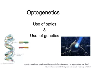
Optogenetics. Genevieve Bell Daniel Blackman Brandon Chelette. Membrane Biophysics - Fall 2014. Optogenetics. Integration of optics and genetics that allows for experimental control of events within a specific cell. Before Optogenetics.

E N D
Optogenetics Genevieve Bell Daniel Blackman Brandon Chelette Membrane Biophysics - Fall 2014
Optogenetics • Integration of optics and genetics that allows for experimental control of events within a specific cell
Before Optogenetics • The spatial and temporal resolution of modulation in the brain left much to be desired • Spatial resolution: stimulation with electrodes does not distinguish between different cell types (Francis Crick, 1979) • Temporal resolution: multi-component systems require a long time to occur • Insufficient for millisecond time scales needed to analyze cellular events such as action potentials
Before Optogenetics • It was known that there were single component, light activated ion pumps in bacteria • Vast array of microbial opsin genes
Optogenetics – the basic concept • Express a light-activated ion channel (isolated from bacteria) in a specific sub-population of neurons • Illuminate neurons to modulate activity • Modulation depends on type of channel • Record output (e-phys, fMRI, behavioral, etc.)
Initial Problems • It was presumed that: • Microbial proteins would not be expressed very well in mammalian cells • Photocurrents not strong enough to control neurons • Retinal (co-factor) would have to be added to the cell type of interest
Initial Problems • It was presumed that: • Microbial proteins would not be expressed very well in mammalian cells (bacterial opsin expressed reliably in mammalian neurons. Boyden 2005) • Photocurrents not strong enough to control neurons • Retinal, a requisite cofactor for opsins, would have to be added to the cell type of interest
Initial Problems • It was presumed that: • Microbial proteins would not be expressed very well in mammalian cells (bacterial opsin expressed reliably in mammalian neurons. Boyden 2005) • Photocurrents not strong enough to control neurons (bacterial opsin provided sustained control of action potential. Boyden 2005) • Retinal, a requisite cofactor for opsins, would have to be added to the cell type of interest
Initial Problems • It was presumed that: • Microbial proteins would not be expressed very well in mammalian cells (bacterial opsin expressed reliably in mammalian neurons. Boyden 2005) • Photocurrents not strong enough to control neurons (bacterial opsin provided sustained control of action potential. Boyden 2005) • Retinal, a requisite cofactor for opsins, would have to be added to the cell type of interest (all vertebrate tissues contain sufficient all-trans retinal. Douglass 2008)
Optogenetics • Allows for high resolution temporal and spatial modulation of the activity of a cell • The population of neurons being targeted, the timing of the modulation, and the nature of the modulation are all under experimental control
Optogenetics: a diverse toolkit • The optogenetic effect experienced by a cell will depend on many factors • The properties of single-component opsin being used • The efficiency of the expression of that opsin • The source/wavelength/intensity of the light • The location and density of the population of neurons being investigated
Optogenetics: a diverse toolkit • Wide variety of opsins
Optogenetics: a diverse toolkit Four main categories of opsins: • Fast excitation • Fast inhibition • Step function • Biochemical modulation
Optogenetics: a diverse toolkit Biochemical modulation • Opsin + G-protein coupled receptor • Activated by light, but not an ion channel • Illumination leads to intracellular signaling cascades • Can be considered slow excitatory or slow inhibitory based on the nature of the G protein signaling pathway • Provide precise control of intracellular signals (cAMP, GTPase, etc.)
Optogenetics: a diverse toolkit Fast excitation • Channelrhodopsins • Cation channels • Modified via mutagenesis with optimization in mind, but can result in unanticipated or undesired effects • Gain-of-function tool
Optogenetics: a diverse toolkit Fast excitation
Optogenetics: a diverse toolkit Fast inhibition • Chloride & proton pumps • Also engineered for optimal function • Loss-of-function tool
Optogenetics: a diverse toolkit Fast inhibition
Optogenetics: a diverse toolkit Step function opsins (SFOs) • Exhibit bistability • Much slower deactivation rate • Product of molecular engineering • Cation channels • Larger disparity between activating wavelength and deactivating wavelength
Optogenetics: a diverse toolkit Step function opsins (SFOs)
Optogenetics – a diverse toolkit • Vast array of opsins available to be used as a result of attempted optimization • Point mutations • Codon insertion/deletion/replacement • Small changes can lead to beneficial or detrimental changes in: • Current generation • Opening/closing kinetics • Sensitization/Desensitization
Optogenetics – targeting your opsin • Viral injection • LV , AAV • Projection targeting • Dendrites or axons • Transgenic targeting • Transgenic mouse lines that are not under recombinase-dependent control • Spatiotemporal targeting • Birthdate of cells , specific layer
Optogenetics – light delivery • Assuming you are expressing the correct opsin in the desired cell population, you now need to somehow get light to those cells • There are several facets to consider and the best choice will depend on your experiment • Excitation vs inhibition vs bistable • Wavelength • Intensity • Duration
Optogenetics – light delivery • Brain tissue scatters and absorbs light • Different tissues scatter and absorb light in different ways (Ex: myelinated tissue scatters light significantly)
Optogenetics – light delivery Different wavelengths of light penetrate brain tissue better than others
Optogenetics – light delivery • Surface targets: cell culture, brain slices, etc • Easily accessible, spot illumination • Deep Targets: in vivo targets that are not on the outer surface of the brain • Minimize damage • Fiber optics
Optogenetics – light sources • Variety of light sources: • Lasers • LEDs • Incandescent • Depending on where you need to deliver your light and how, you need to select the correct light
Optogenetics – the good • Significant experimental control • High resolution temporal and spatial control • Specific cell type population • Millisecond control on time scale of cellular events like action potentials • Wide variety of opsins available • Can be applied to more fields than just neuroscience (cardiac muscle, skeletal muscle, etc) • Potential disease models
Optogenetics – the bad • Damage to surrounding tissue (depending on location of target) • Light absorption heat damage tissue or affect physiological events • Viral infection and/or expression of exogenous proteins can lead to unwanted alterations in cell capacitance, physiological activity, structural abnormalities, and toxicity • Second and third degree currents can confound actual effects • Causality can not always be proven
The ugly take home message • Optogenetics is a burgeoning technique that has provided neuroscientists with a much more finely-tuned way to manipulate the cellular activities of a specific population of cells they are interested in. But with any relatively new technique, there are still kinks that need to worked out
Color-tuned Channelrhodopsins for MultiwavelengthOptogenetics Matthias Prigge, FranziskaSchneider, Satoshi P. Tsunoda, Carrie Shilyansky, Jonas Wietek, Karl Deisseroth, and Peter Hegemann A presentation by Daniel Blackman
Modifying ChRs to Overcome Limitations • Limitations of ChR2s • Low expression • Small conductance • Inappropriate kinetics • Partial inactivation • Ion selectivity • Retinal pocket changes affect absorption, kinetics, and membrane targeting • Glu123Thr/Ala(ChETA variants) • Cys128Ser or Asp156Ala • Glu90, Glu123, Leu132, or His134 • C1 and C2 helix swapping
What Previous Studies Have Taught Us • Volvox C1 (V1) absorption max • Pyramidal neurons • Dual-color activation • Spectrally separated absorption • Large photocurrents • Different operational sensitivities
Spanning the Spectrum • Operational Light Sensitivity • Calcium indicators or voltage sensors • Molecular engineering • Helix Swapping • Global rearrangements
Global Structural Rearrangements Prigge et al (2012)
Imaging and Analysis Prigge et al (2012)
Fast and Slow-Cycling Prigge et al (2012)
AP Firing in Hippocampal Neurons Prigge et al (2012)
Dual Light Excitation and Ion Selectivity Prigge et al (2012)
Current-voltage relationships Prigge et al (2012)
Conclusion • Identified helices H6 & H7 responsible for absorption differences, and helices H1 & H7 for membrane integration • Achieved independent activation of distinct neural populations through AA mutations: • ChR2 with absorption maxima 461-492 nm (blue) • C1V1 chimeric variant with absorption maxima 526-545 nm (green) • C1V1-B & C2-LC-TC provide better expression & lower inactivation • Further color tuning possible through exploring further AA mutations
Fast-conducting mechanoreceptors contribute to withdrawal behavior in normal and nerve injured ratsRirie DG, et al. 2014. Pain. Membrane Biophysics – Fall 2014
Background - Pain • Transduction of noxious (or potentially noxious) stimuli from periphery to central nervous system • Complex integration with emotional and cognitive signals perception of pain
Background - Pain A-fibers – “first pain” C-fibers – “second pain” Julius & Basbaum 2001
Background - Pain Ablation of C-fibers does not eliminate pain behavior Other afferents might be responsible for at least some portion of ongoing pain
Background - Experiment • Neurons of interest: fast-conducting, myelinated, nociceptive, high-threshold mechanoreceptors = AHTMRs • Investigate: what is the role of these neurons in pain signal transduction under normal conditions and under neuropathic conditions?
Background - Experiment • Selectively inhibit these specific neurons (AMHTRs) using expression of light-activated proton pump • Male Sprague-Dawley rats: intrathecal injection of AAV8 with CAG/ArchT/GFP tag
Light activates proton pump Pumps protons intra extra Hyperpolarizes cell / Reduce excitability / Inhibit activity
First things first… Expression of GFP-ArchT in soma, dendrites and axons Expression maximized around 4 weeks post-injection
NF200: marker for myelinated cells IB4: marker for unmyelinated cells