Download
1 / 26
280 likes | 1.71k Views
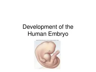
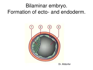
Folding of the Embryo Formation of Gut Endodermal Derivatives. By: Dr. Mujahid Khan. Folding Of Embryo. Flat trilaminar disc folds into a somewhat cylindrical embryo Folding occurs in both median & horizontal planes Results from rapid growth of the embryo

E N D
Folding of the EmbryoFormation of GutEndodermal Derivatives By: Dr. Mujahid Khan
Folding Of Embryo • Flat trilaminar disc folds into a somewhat cylindrical embryo • Folding occurs in both median & horizontal planes • Results from rapid growth of the embryo • Long axis increases rapidly than the sides • Occurs simultaneously on both axis • Constriction at the junction of embryo & yolk sac
Folding in Median Plane • Occurs in the cranial and caudal ends • Causing head and tail folds • Moving ventrally as the embryo elongates cranially and caudally
Head Fold • At the beginning of the 4th week • Neural folds in the cranial region thickened to form primordium of the brain • Initially the developing brain projects dorsally into the amniotic cavity • Later grows cranially beyond the oropharyngeal membrane • Overhangs the developing heart
Head Fold • Septum transversum, primordial heart, pericardial coelom & oropharyngeal membrane move onto the ventral surface • Endoderm of the yolk sac is incorporated into the embryo as a foregut • The foregut lies between the brain & heart • Oropharyngeal membrane separates the foregut from the stomodeum
Head Fold • Septum transversum lies caudal to heart after the folding and develops into central tendon of diaphragm • Head fold also affects the arrangement of the primordium of body cavity which consists of a flattened horseshoe shaped cavity before folding
Tail Fold • Results primarily from growth of the distal part of the neural tube • This is primordium of the spinal cord • As embryo grows, the caudal eminence projects over the cloacal membrane • During folding, part of endoderm is incorporated into the embryo as a hindgut
Tail Fold • Terminal part of the hindgut soon dilates to form the cloaca • Cloaca is the primordium of urinary bladder and rectum • Before folding primitive streak lies cranial to the cloacal membrane • After folding it lies caudal to it
After Tail Fold • The connecting stalk (primordium of umbilical cord) is attached to the ventral surface of the embryo • Allantois (a diverticulum of yolk sac) is partially incorporated into the embryo
Folding in Horizontal Plane • Folding on sides of the embryo produces right and left lateral folds • Is produced by rapidly growing spinal cord and somites • Ventrolateral rolling of the edges of embryonic disc form roughly cylindrical embryo
Folding in Horizontal Plane • As the abdominal walls form, part of endoderm is incorporated into the embryo as the midgut • Initially there is a wide connection between midgut & yolk sac • After folding the connection is reduced to yolk stalk
Folding in Horizontal Plane • Umbilical cord forms from the connecting stalk • As it forms, ventral fusion of the lateral folds reduces the region of communication between intraembryonic and extraembryonic coelomic cavities to a narrow communication • Amniotic cavity expands and obliterates extraembryonic coelom
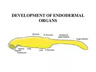
Derivatives of Endoderm Endoderm gives rise to the epithelial lining of: • Trachea • Bronchi • lungs
Derivatives of Endoderm Endoderm gives rise to the epithelial lining of: • Gastrointestinal tract • Liver • Pancreas • Urinary bladder • urachus
Derivatives of Endoderm Endoderm gives rise to the epithelial lining of: • Pharynx • Thyroid • Tympanic cavity • Pharyngotympanic tube • Tonsils • Parathyroid glands
Formation of Gut • Primordial gut at the beginning of the 4th week is closed at its: • Cranial end by oropharyngeal membrane • Caudal end by the cloacal membrane
Formation of Gut • Primordial gut forms during the 4th week as the head, tail and lateral fold incorporate the dorsal part of the yolk sac into the embryo • The endoderm of the primordial gut gives rise to most of the epithelium and glands of the digestive tract
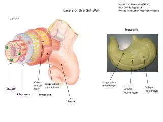
Formation of Gut • The epithelium at the cranial and caudal ends of the tract is derived from ectoderm of the stomodeum (mouth) proctodeum (anal pit) • The muscular, connective tissue, and other layers of the wall of the digestive tract are derived from the splanchnic mesenchyme surrounding the primordial gut
Formation of Gut For descriptive purposes the primordial gut is divided into 3 parts: • Foregut • Midgut • Hindgut