Download
1 / 43
480 likes | 785 Views
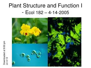
Anatomy, Morphology, & Growth of Angiosperms – Ch. 5-8. Two plant groups: monocots & dicots. Muscle cell. Cells. Tissues. Muscle tissue. Organs. Heart. Circulatory system. Systems. The Plant Cell. Fig 7.8. 5 Differentiated Plant Cell Categories. Parenchyma Collenchyma Schlerenchyma

E N D
Muscle cell Cells Tissues Muscle tissue Organs Heart Circulatory system Systems
The Plant Cell Fig 7.8
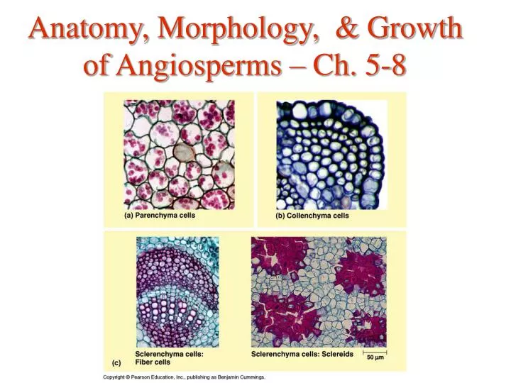
5 Differentiated Plant Cell Categories • Parenchyma • Collenchyma • Schlerenchyma • Water-conducting cells of the xylem • Sugar-conducting cells of the phloem
1. Parenchyma • thin and flexible cell walls
2. Collenchyma • Usually grouped in strands to support young parts of plants without restraining growth • Flexible, elongate with growing shoots
3. Schlerenchyma • May be dead at functional maturity – ??? • cell walls left behind as skeleton
4. Water conducting cells of the xylem: • 2 types: tracheids & vessel elements
Tracheids • Water flows from cell to cell (laterally) through pits in cell wall • Support function
Vessel Elements • End walls are perforated for free flow of water • More efficient as water conductors than tracheids
5. Sugar-conducting cells of the phloem Sieve-tube members: • Lack a nucleus, ribosomes, vacuole • Cells separated by perforated sieve plates – allow sugar movement
Dermal tissue or epidermis • Root hairs are specialized epidermal extensions • Secretes waxy cuticle of the leaf
2. Ground Tissue • fills the space between dermal and vascular tissue systems. • Diverse functions: pith In dicots: cortex
3. Vascular Tissue • function in transport between roots & shoots, and structural support of plant • Xylem: • Phloem: Food transported to roots & non-photosynthetic parts such as the flowers
Growth & Development http://www.cneccc.edu.hk/subjects/bio/album/Chapter20/PLANT_GROWTH.html
Three processes of development: • Growth = • Cellular differentiation = generation of different cell types • Morphogenesis – creation of body form & organization.
1. Growth • Cell division no expansion
Growth • = due to water uptake in the vacuole
Cell division • Occurs in only in meristems!
Meristems • = • Two types of meristems: • Apical meristem – • Lateral meristems – extend lengthwise along the axis of the stem & roots. Responsible for growth in girth in older parts of the plant (called secondary growth). Exist only in perennials
Arrangement of Primary Tissues in Roots • Epidermis – • Stele – • Ground tissue – mostly parenchyma cells of the cortex – area between the stele & epidermis; stores food & takes up minerals. • Endodermis – single cell layer between cortex & stele. Selective barrier for uptake of soil solution contents into vascular system.
Eudicot/Gymnosperm root cross section Epidermis Endodermis Cortex Stele xylem phloem Fig 35.13
Primary Growth of Shoots • Bud = cluster of leaf primordia created by meristem. No internodes • Lateral branches arise from axillary buds
Primary tissue arrangement of stems • Ground tissue = pith & cortex
Eudicot/Gymnosperm stem cross section pith phloem cortex xylem epidermis Fig 35.16 Schlerenchyma cells
Tissue arrangement of leaves • 3 parts: • Upper & lower epidermis – tightly interlocked cells, secrete waxy cuticle. Contains stomata flanked by guard cells • Vascular tissue – • Mesophyll – ground tissue between upper & lower epidermis
Secondary Growth • Two lateral meristems: • Vascular cambium – produces secondary xylem (= wood) & phloem • Cork cambium – replaces the epidermis with cork: tough, thick cover for stems, roots.
Secondary growth of stems • Vascular cambium – layer of cells between primary xylem & primary phloem. Puts on successive layers of secondary phloem to outside & secondary xylem to inside =====> stem widens • Wood = accumulation of secondary xylem. Dead at maturity, contains lignin
What is bark? • “bark” = • Cork continually sloughs off
Three types of life cycles: • Annual – • Biennial – complete life cycle in two years (first year = vegetative, second year = reproductive). Some need a cold winter period to initiate flowering from vegetative state. Ex. carrots • Perennial – live year after year, do not die after reproduction. Examples: trees, shrubs, some grasses. Causes of death = fire, disease