Download
1 / 17
350 likes | 1.46k Views
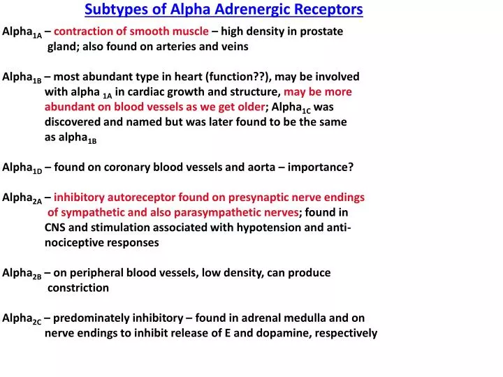
Subtypes of Alpha Adrenergic Receptors. Alpha 1A – contraction of smooth muscle – high density in prostate gland; also found on arteries and veins Alpha 1B – most abundant type in heart (function??), may be involved

E N D
Subtypes of Alpha Adrenergic Receptors Alpha1A – contraction of smooth muscle – high density in prostate gland; also found on arteries and veins Alpha1B – most abundant type in heart (function??), may be involved with alpha 1A in cardiac growth and structure, may be more abundant on blood vessels as we get older; Alpha1C was discovered and named but was later found to be the same as alpha1B Alpha1D – found on coronary blood vessels and aorta – importance? Alpha2A – inhibitory autoreceptor found on presynaptic nerve endings of sympathetic and also parasympathetic nerves; found in CNS and stimulation associated with hypotension and anti- nociceptive responses Alpha2B – on peripheral blood vessels, low density, can produce constriction Alpha2C – predominately inhibitory – found in adrenal medulla and on nerve endings to inhibit release of E and dopamine, respectively
Intrinsic Mechanisms Produced By Receptor Activation Muscarinic 2 receptors: Gi/Go – inhibits adenylyl cyclase, inactivates calcium channels, increases potassium efflux – hyperpolarization INHIBITORY Muscarinic 3 receptors: Gq/11 protein – increase phopholipase C activity, increase formation of IP3 and DAG, increase intracellular calcium CONTRACTION (in most cells – exception – vascular smooth muscle cells) Alpha one receptors: Gq/11 protein – same as muscarinic 3 receptor mechanism - CONTRACTION Alpha 2 receptors: Gi/Go protein – same as muscarinic 2 receptor mechanism – INHIBITORY Beta one receptors: Gs proteins – increase activity of adenylyl cyclase, increase intracellular calcium – EXCITATORY Beta 2 receptors: Gs proteins – increase activity of adenylyl cyclase activity in most smooth muscle cells, decrease intracellular calcium INHIBITORY
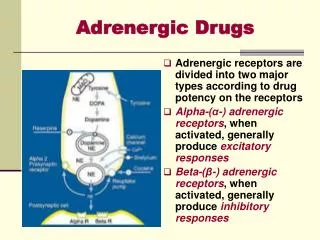
CLASSIFICATION OF RECEPTORS Adrenergic Receptors (all are GPCRs) Dr. Raymond Alquist - 1948 Alpha one receptors – vascular and nonvascular smooth muscle, Gq protein – contraction Alpha two receptors – presynaptic nerve terminals, pancreatic beta cells, vascular smooth muscle, Gi/Go protein – inhibitory most of the time (exception on vascular smooth muscle) Beta one receptors – heart, J-G cells within kidneys, Gs proteins – excitatory Beta two receptors – smooth muscle (vascular, bronchial, GI and UT), Gs protein – inhibitory Beta three receptors – adipose tissue, Gs protein – lipolysis
Receptors at Neuroeffector Junction Involuntary Contraction Of Cardiac Cell Ca++ Ca++ Sarcoplasmic Reticulum Increased Contraction Voltage-gated Channel Depolarization of Cell Ca++ Cardiac Cell
M2 receptor Ca++ adenyl cyclase Sarcoplasmic Reticulum Decreased Contraction or Relaxation ACh Inactivates channel inhibits Gi or oprotein K+ AC – open calcium channel PKA – opens calcium channel and releases Ca++ from SR ATP cAMP Hyperpolarization Active Protein Kinase A Inactive Protein Kinase A Cardiac Cell
Sarcoplasmic Reticulum STIMULI Voltage-gated channel Calmodulin On Myosin Ca++ Ca++ Calmodulin Complex MLCK MLCK* ATP Myosin Light Chain Myosin Light Chain – PO4 Myosin Phosphatase Actin Myosin RELAXATION CONTRACTION Smooth Muscle Cell
PLC Sarcoplasmic Reticulum ACh PIP2 M3 Receptor DAG Gq Protein Ca++ IP3 Ca++ Protein Kinase C Calmodulin ATP ADP Calmodulin Complex PO4 MLCK MLCK* Myosin Light Chain Myosin Light Chain – PO4 Actin CONTRACTION Smooth Muscle Cell PIP2 = phosphatidyl inositol biphosphate IP3 = Inositol triphosphate DAG = Diaacylglycerol
Acetylcholine PLC Gq Protein eNOS Nitric Oxide Sarcoplasmic Reticulum Muscarinic 3 Receptor PIP2 IP3 L-Arginine Ca++ Calmodulin Ca++-Calmodulin Complex L-Citrulline Endothelial Cell Lining Blood Vessel Lumen
PLC R E L A X A T I O N Nitric Oxide Calmodulin Guanyl Calmodulin Complex Cyclase MLCK* MLCK Sarcoplasmic Reticulum Myosin Light Chain Actin CONTRACTION Muscarinic 3 Receptor Ca++ Ca++ Cyclic GMP GTP inhibits Ca++ Myosin Light Chain Myosin Light Chain – PO4 Myosin Phosphatase Vascular Smooth Muscle Cell
Receptors at Neuroeffector Junction γ β α NE G Protein-Coupled Receptor Second Messenger Receptor Ca++ Effector Protein (Adenyl Cyclase) cAMP ATP GTP GDP GDP 5’AMP Beta receptor PDE RESPONSE
NE PLC Sarcoplasmic Reticulum PIP2 Alpha1 DAG Gq Protein Ca++ IP3 Ca++ Protein Kinase C Calmodulin ATP ADP Calmodulin Complex PO4 MLCK MLCK* Myosin Light Chain Myosin Light Chain – PO4 Actin CONTRACTION Smooth Muscle Cell
Alpha 2 Presynaptic Ca++ adenyl cyclase Alpha 2 Receptor Agonist Inactivates channel inhibits Gi or o protein K+ ATP cAMP Hyperpolarization Decrease Release of Neurotransmitter Presynaptic Nerve Terminal or CNS
Beta-1 Receptor Ca++ Ca++ adenyl cyclase Sarcoplasmic Reticulum Increased Contraction NE Ca++ Gs protein ATP cAMP phosphorylation Active Protein Kinase A Inactive Protein Kinase A Enhance actin and myosin interaction Ca++ Increased Ca++ Binding to troponin Cardiac Cell
Adenyl Cyclase Gs Protein Sarcoplasmic Reticulum Myosin Light Chain – PO4 Myosin Light Chain CONTRACTION Actin RELAXATION Epi., Albuterol Terbutaline Beta Two Receptor cAMP ATP Ca++ act. PKa Ca++ abnormal phosphorylation Calmodulin Calmodulin Complex K+ MLCK MLCK* *(inactive) Hyperpolarizatiion Smooth Muscle Cell
Responses of Effector Organs to Autonomic Nerve Impulses α1 α1 α1 α1 α1 Sympathetic and Parasympathetic TONE Continually active – SNS: Blood Vessels - maintain peripheral resistance PNS: Heart Loss of sympathetic tone increase in intrinsic tone of smooth muscle Denervation Supersensitivity α1 Sympathetic or Parasympathetic stimulation of receptors can result in Excitatory Effects in some organs but Inhibitory Effects in others! Frequently, if sympathetic stimulation causes excitation in an organ, parasympathetic stimulation to that same organ will result in inhibition.