Download
1 / 1
10 likes | 142 Views
No. 053. Change in Size of Cystocele between Supine and Upright Position. Sharon English, Nadya York Christchurch Public Hospital. Introduction

E N D
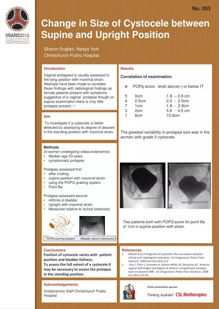
No. 053 Change in Size of Cystocele between Supine and Upright Position Sharon English, Nadya York Christchurch Public Hospital Introduction Vaginal prolapsed is usually assessed in the lying position with maximal strain. Attempts have been made to correlate these findings with radiological findings as female patients present with symptoms suggestive of a vaginal prolapse though on supine examination there is only little prolapse present. 1,2 Results Correlation of examination n POPq score level above(-) or below IT 5 0cm -1.8 – 2.9 cm 4 0.5cm -2.0 - 2.0cm 7 1cm -1.8 - 2.9cm 3 2cm 3.8 - 4.5 cm 1 6cm 10.9cm The greatest variability in prolapse size was in the women with grade II cystocele Aim To investigate if a cystocele is better detected by assessing its degree of descent in the standing position with maximal strain. • Methods • 20women undergoing videourodynamics • Median age 53 years • symptomatic prolapse • Prolapse assessed first • after voiding, • supine position with maximal strain • using the POPQ grading system • Point Ba • Prolapse assessed second • 400mls in bladder • Upright with maximal strain • Measured relative to ischial tuberosity Two patients both with POPQ score for point Ba of 1cm in supine position with strain. POPq scoring system Bladder above tuberosity(-) Conclusions Position of cystocele varies with patient position and bladder fullness. To assess the full extent of a cystocele it may be necessary to assess the prolapse in the standing position. References Altman D et al Diagnosis of cystocele--the correlation between clinical and radiological evaluation. IntUrogynecol J Pelvic Floor Dysfunct. 2004 Jan-Feb;15(1):3-9; Hsu Y, Chen L, Summers A, Ashton-Miller JA, DeLancey JO. Anterior vaginal wall length and degree of anterior compartment prolapse seen on dynamic MRI. IntUrogynecol J Pelvic Floor Dysfunct. 2008 Jan;19(1):137-42. Acknowledgements Urodynamics Staff Christchurch Public Hospital Poster presentation sponsor