Download
1 / 46
470 likes | 773 Views
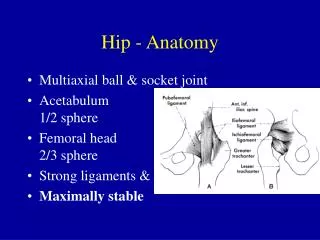
Hip problems. S lipped C apital F emoral E piphysis P erthes D isease D evelopmental D ysplasia of H ip. Slipped Capital Femoral Epiphysis. Introduction. The most common hip abnormality presenting in adolescence and a primary cause of early osteoarthritis.

E N D
Hip problems • Slipped Capital Femoral Epiphysis • PerthesDisease • DevelopmentalDysplasiaof Hip
Introduction • The most common hip abnormality presenting in adolescence and a primary cause of early osteoarthritis. • SCFE is a Salter-Harris type 1 fracture through the proximal femoral physis. • The femoral head gradually slip posteriorly, medially and intefriorly with respect to the neck.
Incidence • Incidence is 1 case per 100,000 people. • SCFE occurs most frequently in adolescents overweight boys African American children • Left hip more common than right. • Bilateral involvement 20-40%
Etiology • Unknown! • Any condition that decreases physeal strength • Increased shear forces due to obesity • ↑weight during growth spurt=↑strain on growth plate • More vertical proximal femoral physis • Retroversion ↑ shear forces with walking • Disease processes that weaken physis • Hypothyroidism • Renal disease • Hypogonadism • Hypopituitarism • GH deficiency treated with HGH • <10 years of age • Other factors that contribute • Marfan’s disease • Posttransplant medications • Radiation therapy
Clinical presentation • Clinical presentation often is misleading, with only 50% of patients presenting with hip pain and 25% presenting with knee pain. • Antalgic limp: Knee pain; 46% • Severe pain= unable to walk • Extremity: externally rotated/adducted/ shortened • Lack of Internal Rotation • Flex hip externally rotate
Symptoms and Clinical Findings • Antalgic limp: Knee pain; 46% • Severe pain= unable to walk • Extremity: externally rotated/adducted/ shortened • Lack of Internal Rotation • Flex hip externally rotate
Radiology • Diagnosis is made using AP pelvis and lateral frog-leg radiographs. • Abduction of the femur for the frog-leg view may result in increased slippage and should be performed with caution.
Radiographs • AP • May not detect the slip! • Widening early dz • Irregular growth plate • Steel’s metaphyseal blanching • No remodeling: acute unstable • Change in Klein’s line Line is not intersecting any part of the head
Radiology Signs • Loss of triangular sign of capener • Blurring of physis • Relative decreased height of epiphysis • Loss of intersection of epiphysis by lateral cortical line of femoral neck.
Radiographs • Head-Shaft angle by Southwick, • Determine degree of slip/stability on frog-leg lateral • Angle between femoral head and shaft (HSA) • HSA of affected side minus HSA of nl side • Determine long-term px • Mild: 1-29 • Mod: 30-60 • Severe: >60 • CT Scan • Assess magnitude of deformity • Post-op check of physeal closure • MRI • Dx of AVN or chrondrolysis
13-year-old female adolescent • anteroposterior pelvic view: increased opacity of her right metaphysis and the subtle widening of the physis
14-year-old male adolescent who came to the emergency department with complaints of thigh and knee pain • A relatively subtle medial slip at AP view. • A more obvious posterior slip at frog-leg lateral view
Classification • Traditional Classification • Fahey and O’Brian, 1965 • no rationale for this selection of time • Acute < 3 weeks of symptoms • Chronic > 3 weeks of symptoms • Acute on Chronic > 3 weeks of symptoms + sudden exacerbation
Surgical Treatment • Goals: • “primum non nocere” • Pain relief • Prevent slip progression • Accelerate epiphsiodesis • Avoid AVN and chondrolysis
Pinning In-Situ • Single-screw in-situ • High success rate • Low incidence of slippage • Minimal complications with proper placement • Placement of single screw • 1980’s: Morrissey • Center of femoral head/┬ to physis • Enhance rate of physeal closure (Ward, JBJS, 1992) • Avoid posterosuperior quadrant (Brodetti, JBJS, 1960) • Injury to lateral epiphyseal vessels= AVN
Pinning In-Situ • Placement of single screw • Proper start point important • anterior on neck • pin start below lesser troch: ↑ fx incidence • Multiple drill holes weaken bone • 5 screw threads into the epiphysis
Pre-Op Screw Placement
2-Bone-Graft Epiphysiodesis 3-Osteotomy
Complication • AVN • Chondrolysis • Continued Slip • Because of smooth pins • Poor primary fixation: not perpendicular to physis • Pin Breakage: unstable fixation • Subtrochanteric fracture • OA/Pistol grip deformity
Perthes Disease LEGG-CALVE-PERTHES DISEASE
Perthes Disease • Idiopathic Avascular Necrosis of Capital Femoral Epiphysis (CFE) • An ischaemic episode of the lateral epiphyseal arteries initiates avascular necrosis of the capital femoral epiphysis. The lateral epiphyseal arteries supply an extensive area of the capital femoral epiphysis. • Cause is unknown
Perthes Disease • More common in boys than girls 4-5:1 • Age range 3-11yo more usually 5-6 yo • Often lower socio-economic groups (?nutrition factors) Perthes in Lt. femoral head
Perthes Disease Etiology • interruption of blood supply to CFE • ossification ceases temporarily • articular cartilage continues to grow (nourished by synovial fluid) • subchondral bone is revascularised and becomes weak • results in subchondral fracture which can allow the femoral head to become flattened and mis-shapen
Perthes Disease Signs & Symptoms • Limp • Pain in knee, thigh • ↓ hip ROM abduction & internal rotation • Affected leg may become shorter and thinner over time
Perthes Disease • Physical Exam - Shows • 1. Decrease ROM in hip abduction and internal rotation. • 2. Hip stiffness • 3. Knee pain • X-rays: Four stages • 1. Synovitis • 2. Aseptic necrosis- increased joint space and small femoral head • 3. Fragmentation - increased bone density • 4. Residual - increased bone density
Perthes Disease Management: • Many (approx 60%) do well without treatment of any kind, especially younger boys under 5 years of age. • Some (approx. 15%) do badly even with active management. • Remaining 25% benefit from active management. • Factors that determine which group children will fall into has been difficult to determine. Following principles are generally agreed. Management Principles • reduce hip irritability, pain and spasm if present • prevent deformity of the femoral head (reduces risk for osteoarthritis in later years) • Congruity of hip joint.
Perthes Disease • Rest in bed with pain relief • Traction to relieve muscle spasm • Slings/springs to regain ROM • Containment of femoral head in acetabulum through • use of abduction brace (eg. Scottish Rite or Toronto). Continue to ambulate. • surgically increasing acetabular coverage (innominate and/or varus osteotomy) followed by period in broomstick plasters.
Perthes Disease • Avoid high impact activities eg running, jumping until fem. head is healed. • Hydrotherapy may also be useful. • Recovery is a slow process (2-5 years) therefore chn. need emotional support and reassurance that they will recover and be able to resume "normal" activities.
DevelopmentalDysplasiaof theHip • Femoral head has an abnormal relationship with acetabulum • Includes unstable, subluxated (excessive movement in the socket), and dislocated hips • Risk Factors • Female • Breech • Family History
DDH • Recommendations • Serial physical screening exams • Hip imaging for females born breech (120/1000) • Optional imaging for males born breech (26/1000) • Optional imaging for females with FH • If positive Ortalani or Barlow on initial PE, refer to orthopedic doctor • If exam is “equivocal” (soft clicks)check in 2 wks
DDH • DDH • Developmental Dysplasia of the Hip • CDH • Congenital Dislocation of the Hip
Radiological Diagnosis • classic features • increased acetabular index ( n=27, >30-35 dysplasia) • disruption shenton line ( after age 3-4 should be intact on all views) • absent tear drop sign • delayed appearance ossific nucleus and decreased femoral head coverage • failure medial metaphyseal beak of proximal femur , secondary ossification center to be located in lower inner quadrant • center-edge angle useful after age 5 ( < 20) when can see ossific nucleus
DDH Physical findings • Girl • Asymmetrical skin folds • Limited abduction • Short leg • Pistoning • Ortolani’s sign • Barlow’s sign
DDH X-ray findings • Delayed appearance of ossific nucleus • Small ossific nucleus • Dysplastic acetabulum • Proximal displacement of femur
DDH Treatment • 0 – ½: Pavlik harness • ½ – 1½: Closed reduction, cast • 1 ½ - 5 or 8: Open reduction, pelvic osteotomy • Older: Leave dislocated • Pavlik Harness • Check at 3 weeks to confirm reduction • Adjust position every 6 – 12 weeks • Continue until the hips are clincally and radiologically normal
Management of DDH • Newborn • Splintage in abduction (Pavlik harness) • 6 - 18 months • Closed reduction - Traction • Splintage • Open reduction and Splintage • Late diagnosed dislocations • Persistent dislocation in adults
Treatment of DDH • Group I - Neonate to 6 weeks - positive Ortolani and Barlow’s tests and skin fold discrepancies. Also dislocated side can be extended all the way down to the level of the exam table, because it is lacking the normal hip flexion tightness that newborn have. Refer this child to Orthopedics for treatment most likely with a Pavlik harness.
Treatment of DDH • Group II - 6 weeks - 12 months - Hip capsular and soft tissue have now tightness up and the Ortolani test may not be positive. Will see limited abduction in this age and skin fold asymmetry. Again referral to Ortho for treatment with Pavlik harness, traction, adductor tenotomy, or closed reduction.
Treatment of DDH • Group III - 12 months - 3 years - Walking with a painless limp. Galeazzi sign positive, and limited abduction. X-rays positive by this age. Again referral to Ortho for possible treatment by arthrography, traction, adductor tenotomy, open reduction, and pelvic versus femoral osteotomy.
Treatment of DDH • Group IV - 3 years to skeletal maturity- Same as group III and X-ray is positive. Referral to Ortho for treatment. Usually need to have surgery to corrected at this age. • FYI - Bilateral dislocations over 6 years old and unilateral over 8 years old do better left ALONE.