Download
1 / 44
520 likes | 1.01k Views
Interpretation of laboratory Tests General & Cardiovascular System. Hadeel Alkofide MSc PHCL 326 May 2011. Learning Objectives. Differentiate between invasive & noninvasive tests State the clinical application of common general diagnostic procedures

E N D
Interpretation of laboratory TestsGeneral & Cardiovascular System Hadeel Alkofide MSc PHCL 326 May 2011
Learning Objectives • Differentiate between invasive & noninvasive tests • State the clinical application of common general diagnostic procedures • Identify the clinical application of specific laboratory tests • Identify the clinical application of specific diagnostic procedures • Assess common laboratory & diagnostic test results
Introduction • Data from laboratory & diagnostic tests & procedures provide important information regarding • The response to drug therapy • The ability of patients to metabolize & eliminate specific therapeutic agents • The diagnosis of disease, & the progression & regression of disease
Introduction Invasive tests • Require penetration of the skin or insertion of instruments or devices into a body orifice • The degree of risk varies from relatively minor risks such as pain, bleeding, & bruising associated with venipuncture to the risk of death associated with more invasive procedures such as coronary angiography • E.g venipuncture, insertion of a central venous catheter, & collection of cerebrospinal fluid
Introduction Noninvasive tests • Do not penetrate the skin or involve insertion of instruments into body orifices & pose little risk to the patient • E.g. include chest radiograph, analysis of spontaneously voided urine, & stool occult blood analysis
Introduction • The selection of specific tests & procedures depends on • The patient's underlying condition • The need for the information • The degree of risk • Reference ranges are listed in Tables 5-1 through 5-7 • Results are interpreted using laboratory specific reference ranges • Reference ranges may differ among different laboratories depending on the population & laboratory methodology used to establish the range
General Organ System Monitoring • A variety of tests & procedures are used to diagnose & monitor conditions that affect various organ systems • The applications & uses of these tests & procedures continue to expand with experience & the integration of new technology
General Organ System Monitoring Angiography • A radiographic test used to evaluate blood vessels & the circulation • Radiopaque material is injected through a catheter & images are recorded using standard radiographic techniques Biopsy • Removal & evaluation of tissue
General Organ System Monitoring Angiography
General Organ System Monitoring Computed Tomography • (CT; CAT scan) uses a computerized X-ray system to produce detailed sectional images • The system is very sensitive to differences in tissue density & produces detailed, two-dimensional planar images • Contrast agents increase attenuation • The spiral or helical CT takes pictures continuously, decreasing the time needed to obtain images
General Organ System Monitoring Computed Tomography
General Organ System Monitoring Doppler Echography • Uses ultrasound technology to measure shifts in frequency from moving images • E.g, Doppler echography is used to evaluate blood flow velocity & turbulence in the heart (Doppler echocardiography) & peripheral circulation
General Organ System Monitoring Doppler Echography
General Organ System Monitoring Endoscopy • Used to examine the interior of a hollow viscus (e.g., digestive, respiratory, & urogenital organs & the endocrine system) or canal (e.g., bile ducts, pancreas) • The endoscope, a flexible or inflexible tube with a camera & a light source, is inserted into a body orifice (Figure5-1) • Still &/or video images are recorded & tissues obtained for biopsy or other laboratory diagnostic tests
General Organ System Monitoring Endoscopy: Examples of common endoscopic procedures • Colonoscopy: views the inside of the entire colon from rectum to end of the small intestine • Sigmoidoscopy: views the inside of the large intestine from the rectum through the sigmoid colon • Cholangiopancreatography: views the inside of the bile ducts & pancreas • Esophagogastroduodenoscopy: views the inside of the esophagus, stomach, & duodenum • Bronchoscopy: views the inside of the tracheobronchial tree
General Organ System Monitoring Fluoroscopy • Uses a fluoroscope, a device that makes the shadows of x-ray films visible, to provide real-time visualization of procedures • It exposes a patient to more radiation than routine radiography but often is used to guide needle biopsy procedures & nasogastric tube advancement
General Organ System Monitoring Magnetic Resonance Imaging • Uses an externally applied magnetic field to align the axis of nuclear spin of cellular nuclei • The patient is surrounded by the magnetic field (Fig. 5-2) • Brief radio frequency pulses are applied to displace the alignment • The energy emitted when the displacement ends is detected resulting in finely detailed planar & three-dimensional images • Contrast agents increase the attenuation
General Organ System Monitoring Magnetic Resonance Imaging
General Organ System Monitoring Paracentesis • The removal & analysis of fluid from a body cavity Plethysmography • Measures changes in the size of vessels & hollow organs by measuring displacement of air or fluid from a containment system • Body plethysmography is used to assess pulmonary function
General Organ System Monitoring Plethysmography
General Organ System Monitoring Positron Emission Tomography (PET) • Uses positron-emitting radionuelides to visualize organs & tissues of the body • The radionuclides decay, producing positrons that collide with electrons • A special camera detects photons, released when the positrons & electrons collide • It provides quantitative information regarding the structure & function of organs & tissues
General Organ System Monitoring Positron Emission Tomography (PET)
General Organ System Monitoring Single-Photon Emission Computed Tomography. (SPECT) • Similar to PET but involves the administration of radionuclides that emit gamma rays • It is less expensive than PET but provides limited image resolution
General Organ System Monitoring Standard Radiography (Plain Films, X-Ray Films) • Produces images on photographic plates (Figure 5-3) • These films are sometimes difficult to interpret because the three dimensionality is lost on the planar images
General Organ System Monitoring Ultrasonography (Echography) • Uses ultrasound (high-frequency waves imperceptible to the human ear) to create images of organs & vessels • Ultrasonography is used to visualize the fetus in uterus
Cardiovascular Laboratory Tests • Cardiac Enzymes • The pattern & time course of the appearance of enzymes in the blood after cardiac muscle cell damage are used to diagnose MI • CreatineKinase • Lactic dehydrogenase (LDH) • Troponins
Cardiovascular Laboratory Tests Cardiac Enzymes • CreatineKinase • Found in skeletal & cardiac muscle, brain, bladder, stomach, & colon • Isoenzyme fractions identify the type of tissue damaged • CK-MB(CK2) is found in cardiac tissue • CK-MB is detected in the blood within 3 to 5 hours after a MI • Levels peak in about 10 to 20 hrs & normalize within about 3 days
Cardiovascular Laboratory Tests Cardiac Enzymes • Lactic dehydrogenase (LDH) • Found in a variety body tissues • Isoenzyme fractions used to identify type of tissue damage • LDH1- LDH2 are found in the heart, brain, & erythrocytes • LDH 2 accounts for the highest % of total serum LDH • After a MI the rise in LDH 1 > the rise in LDH2 • LDH ↑ within 12 hrs after an MI • It Peaks 24 to 48 hrs, & normalizes by about-day 10
Cardiovascular Laboratory Tests Cardiac Enzymes • Troponins • A complex of proteins (Troponin I, C, & T) that mediate the actin & myosin interaction in muscle • Troponins I & T are specific to cardiac muscle & are used to identify cardiac muscle injury • Their concentrations ↑ within a few hrs of cardiac muscle injury & remain elevated for 5-7 days
Cardiovascular Laboratory Tests • Cardiac enzymes: CK-MB = Creatinine Kinase-MyoGlobin
Cardiovascular Laboratory Tests • Cholesterol • Low-density lipoprotein (LDL)is strongly correlated with coronary artery disease • High density lipoprotein (HDL)is inversely correlated with coronary artery disease • Triglycerides • Found in very low density lipoproteins (VLDLs) & chylomicrons
Cardiovascular Laboratory Tests • C-Reactive Protein • It is a biologic marker of systemic inflammation. • Preliminary studies have linked an ↑ C-reactive protein with an ↑ risk of MI, stroke, & peripheral arterial disease • Myoglobin. • A small protein found in cardiac & skeletal muscle • The presence of myoglobin in the urine or plasma is a relatively sensitive indicator of cellular damage
Cardiovascular Diagnostic Tests & Procedures • Cardiac Catheterization • Used to evaluate cardiac function • A catheter is passed into the right or left side of the heart • Right-sided catheterization is used to measure right atrial pressures, right ventricular pressures, pulmonary artery pressures, & pulmonary artery occlusion pressure • Left-sided catheterization is used to measure left ventricular pressures
Cardiovascular Diagnostic Tests & Procedures • Chest Radiography. • Chest x-ray films are used to diagnose cardiac disease & monitor the patient's response to drug & nondrug therapy • The chest radiograph is used to determine the size & shape of the atria & ventricles, to calculate the cardiothoracic ratio, & to detect abnormalities in the lung fields & pleural spaces • Coronary Angiography • In coronary angiography the cardiac vessels are visualized by injecting the vessel with a contrast agent
Cardiovascular Diagnostic Tests & Procedures • Echocardiography • Used to evaluate the size, shape, & motion of the valves, & walls & changes in chamber size during the cardiac cycle
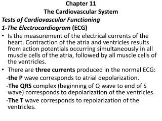
Cardiovascular Diagnostic Tests & Procedures • Electrocardiogram (ECG) • Records the electrical activity of the heart (Figure 5-5) • used to diagnose cardiac disease, monitor the patient's response to drug therapy, & monitor for adverse drug effects • 12 separate leads, 6 extremity (limb) leads & 6 chest leads create a three-dimensional view of cardiac electrical activity
Cardiovascular Diagnostic Tests & Procedures • Electrocardiogram with Stress (Stress Test) • The ECG is recorded during standardized exercise protocol with gradually increasing level of exercise or with patient at rest after administration of dobutamine or dipyridamole • Either intervention increases myocardial oxygen consumption and blood flow • BP, HR, O2 consumption, O2 saturation, & arterial blood gases are collected to provide a thorough assessment of how the CV system functions under stress conditions
Cardiovascular Diagnostic Tests & Procedures • Electrocardiography • Holter Monitoring (Ambulatory Electrocardiography) • A portable recorder to record the ECG continuously throughout usual patient activity • Thallium Stress Test • It combines the parenteral administration of thallium-201, a radionuclide taken up by healthy myocardial tissue, and the stress test (either exercise or pharmacologic) • A gamma camera is used to record serial images of the myocardium
Cardiovascular Diagnostic Tests & Procedures • Technetium-99m Pyrophosphate Uptake. • Infarcted myocardial tissue has an increased uptake of technetium-99m compared with healthy tissue • The isotope is injected parenterally, & serial images of the heart are obtained to evaluate the location & extent of the myocardial infarction