Download
1 / 1
10 likes | 185 Views
Meprin B degradation of tight junction proteins in kidney cells subjected to hypoxia. Non-transfected. Meprin α -transfected. Meprin β -transfected. Non-transfected. Meprin β transfected. E-cadherin. Non-transfected control cells. Meprin β transfected cells. HIF1 α. HIF1 α.

E N D
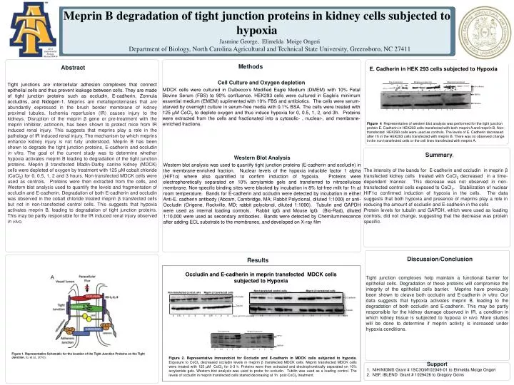
Meprin B degradation of tight junction proteins in kidney cells subjected to hypoxia Non-transfected Meprin α-transfected Meprin β-transfected Non-transfected Meprin β transfected E-cadherin Non-transfected control cells Meprin β transfected cells HIF1α HIF1α E-Cadherin Jasmine George, Elimelda Moige Ongeri Department of Biology, North Carolina Agricultural and Technical State University, Greensboro, NC 27411 GAPDH Tubulin 0 0.5 1 2 3 0 0.5 1 2 3 h Tubulin 2013 Experimental Biology, Abstract #5419 0 1 2 3 0 1 2 3 0 1 2 3 h 0 0.5 1 2 3 0 0.5 1 2 3 hours Methods Abstract E. Cadherin in HEK 293 cells subjected to Hypoxia Cell Culture and Oxygen depletion MDCK cells were cultured in Dulbecco’s Modified Eagle Medium (DMEM) with 10% Fetal Bovine Serum (FBS) to 90% confluence. HEK293 cells were cultured in Eagle’s minimum essential medium (EMEM) suplimented with 10% FBS and antibiotics. The cells were serum-starved by overnight culture in serum-free media with 0.1% BSA. The cells were treated with 125 µM CoCl2 to deplete oxygen and thus induce hypoxia for 0, 0.5, 1, 2, and 3h. Proteins were extracted from the cells and fractionated into a cytosolic- , nuclear-, and membrane-enriched fractions. Tight junctions are intercellular adhesion complexes that connect epithelial cells and thus prevent leakage between cells. They are made of tight junction proteins such as occludin, E-cadherin, Zonnula occludins, and Nidogen-1. Meprins are metalloproteinases that are abundantly expressed in the brush border membrane of kidney proximal tubules. Ischemia reperfusion (IR) causes injury to the kidneys. Disruption of the meprin β gene or pre-treatment with the meprin inhibitor, actinonin, has been shown to protect mice from IR induced renal injury. This suggests that meprins play a role in the pathology of IR induced renal injury. The mechanism by which meprins enhance kidney injury is not fully understood. Meprin B has been shown to degrade the tight junction proteins, E-cadherin and occludin in vitro. The goal of the current study was to determine whether hypoxia activates meprin B leading to degradation of the tight junction proteins. Meprin β transfected Madin-Darby canine kidney (MDCK) cells were depleted of oxygen by treatment with 125 μM cobalt chloride (CoCl2) for 0, 0.5, 1, 2 and 3 hours. Non-transfected MDCK cells were used as controls. Proteins were then extracted from the cells, and Western blot analysis used to quantify the levels and fragmentation of occludin and E-cadherin. Degradation of both E-cadherin and occludin was observed in the cobalt chloride treated meprin β transfected cells but not in non-transfected control cells. This suggests that hypoxia activates meprin B, leading to degradation of tight junction proteins. This may be partly responsible for the IR induced renal injury observed in vivo. Figure 4 Representative of western blot analysis was performed for the tight junction protein E. Cadherin in HEK293 cells transfected with both meprin A and meprin B. Non-transfected HEK293 cells were used as controls. The levels of E. Cadherin decreased after 1h in the HEK293 cells transfected with meprin B. There was no observed change in the non transfected cells or the cell lines transfected with meprin A. Summaryy Western Blot Analysis Western blot analysis was used to quantify tight junction proteins (E-cadherin and occludin) in the membrane-enriched fraction. Nuclear levels of the hypoxia inducible factor 1 alpha (HIF1α) where also quantified to confirm induction of hypoxia. Proteins were electrophoretically separated on 10% acrylamide gels and transferred to nitrocellulose membrane. Non-specific binding sites were blocked by incubation in 8% fat-free milk for 1h at room temperature. Bands for E-cadherin and occludin were detected by incubation in either Anti-E. cadherin antibody (Abcam, Cambridge, MA; Rabbit Polyclonal, diluted 1:1000) or anti-Occludin (Origene, Rockville, MD; rabbit polyclonal, diluted 1:1000). Tubulin and GAPDH were used as internal loading controls. Rabbit IgG and Mouse IgG (Bio-Rad), diluted 1:10,000 were used as secondary antibodies. Bands were detected by Chemiluminescence after adding ECL substrate to the membranes, and developed on X-ray film The intensity of the bands for E-cadherin and occludin in meprin β transfected kidney cells treated with CoCl2 decreased in a time-dependent manner. This decrease was not observed in non-transfected control cells exposed to CoCl2. Stabilization of nuclear HIF1α confirmed induction of hypoxia in the cells. The data suggests that both hypoxia and presence of meprins play a role in reducing the amount of occludin and E-cadherin in the cells Protein levels for tubulin and GAPDH, which were used as loading controls, did not change, suggesting that the decrease was protein specific. Results Discussion/Conclusion Occludin and E-cadherin in meprin transfected MDCK cells subjected to Hypoxia Tight junction complexes help maintain a functional barrier for epithelial cells. Degradation of these proteins will compromise the integrity of the epithelial cells barrier. Meprins have previously been shown to cleave both occludin and E-cadherin in vitro. Our data suggests that hypoxia activates meprin B, leading to the degradation of both occludin and E-cadherin. This may be partly responsible for the kidney damage observed in IR, a condition in which kidney tissue is subjected to hypoxia in vivo. More studies will be done to determine if meprin activity is increased under hypoxia conditions. Non-transfected control cells Meprin β transfected cells Occludin Figure 1. Representative Schematic for the location of the Tight Junction Proteins on the Tight Junction. (Li et al., 2013). Tubulin Figure 2. Representative Immunoblot for Occludin and E-cadherin in MDCK cells subjected to hypoxia. Exposure to CoCl2 decreased occludin levels in meprin β transfected MDCK cells. Meprin transfected MDCK cells were treated with 125 µM CoCl2 for 0-3 h. Proteins were then extracted and electrophoretically separated on 10% acrylamide gels. Western blot analysis was used to probe for occludin. Tubilin was used as a loading control. The levels of occludin in meprin transfected cells started decreasing at 1h post-CoCl2 treatment. Support 1. NIH/NIGMS Grant # 1SC3GM102049-01 to Elimelda Moige Ongeri 2. NSF, iBLEND Grant # 1029426 to Gregory Goins 0 0.5 1 2 3 0 0.5 1 2 3 hours post CoCl2 exposure