Download
1 / 29
490 likes | 1.39k Views
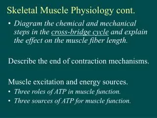
Physiology of Skeletal Muscle Contraction. Dr.Mohammed Alotaibi MRes , PhD (Liverpool, England) Department of Physiology College of Medicine King Saud University. Physiologic Anatomy of Skeletal Muscle.

E N D
Physiology of Skeletal Muscle Contraction Dr.MohammedAlotaibi MRes, PhD (Liverpool, England) Department of Physiology College of Medicine King Saud University
Physiologic Anatomy of Skeletal Muscle • About 50% of the body are muscles: 40% is composed of skeletal and 10% is smooth and cardiac muscles. • Muscle RMP = -90 mV ( same as in nerves ) . • All skeletal muscles are composed of numerous fibers ranging from 10-80 μM in diameter. • Skeletal muscle can be hundreds of centimeters long & is covered by a cell-membrane called Sarcolemma. • The whole muscle is composed of fasciculi»»»»» muscle fiber »»»»» myofibril
Sarcoplasm Matrix inside muscle fiber in which myofilamentssuspended, it is filled with ICF and contains K, Mg, Phosphate, and proteins. Also present are large number of mitochondria to supply ATP. Sarcoplasmic reticulum (SR) Surrounding each myofibril is an extensive membrane-enclosed intracellular compartment called the SR, which plays a key role in activating muscle contraction Transverse tubules (T-tubule) The T-tubule membrane is continuous with the surface membrane.
Each muscle fiber contains between several hundreds to several thousands Myofibrils, the myofibril is striated Dark bands (called A-bands) Light bands (called I-bands) Each Myofibril is made of 3000 Actin filaments and 1500 Myosin filaments .
Each Myofibril contains Actin filaments (thin) & Myosin (thick) filaments which are partially interdigitated. • Each myofibril is striated: consisting of dark bands (called A-bands) and light band (I-bands). Actin Myosin
Sarcomere: Contractile unit of muscle, it is the zone between two Z lines ( discs)= 2 micrometer in length in resting state. Z discs (lines) = lines extend all the way across myofibrils • Inside each sarcomere there are 3 bands: I band = of actin only H band = of myosin only A band = formed of actin & myosin filaments
Muscle Proteins There are four important muscle proteins : • Two contractile proteins that slide upon each other during contraction : • Actin • Myosin • Two regulatory proteins : • Troponin excitatory to contraction • Tropomyosin inhibitory to contraction
Molecular Characteristics of Contractile Filaments Myosin Actin
Myosin (Thick Filament) • Each Myosin molecule has • (1) Head • ( 2 ) Tail • (3) Hinge (joint ) • Furthermore , each myosin head contains • (1) Actin binding site • (2) ATPase site .
- Actin is made of globularprotein callledG-actin- G-actins are attached together to form F-actin strand ( chains ) - Each two strands wind togetherto form double helix called Actin Filament - Tropomyosinlies in the groove between the F-actin strands to cover the active sites on actin that bind the head of myosin- Troponinis attached to tropomyosin and to actin Actin (Thin Filament) G-actin F-actin strand
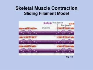
Sliding Filament Mechanism When contraction takes place: Actin & Myosin slide upon each other & the distance between two Z-lines decreases. This is called Sliding Filament . Z-lines come closer to each other, I-band gets smaller , and eventually may disappear, A-band does not become smaller or bigger
Walk-along Theory • Attachment of Myosin to Actin activates the enzyme ATPase in the Myosin Head ATPasebreaks down ATP releasing energy • This energy is used in the “Power Stroke ” to move the myosin head leading to pulling & dragging of actin sliding of actin on myosin • The “ power stroke ” means tilting of the Myosin cross-bridge and dragging ( pulling ) of actin filament
Excitation of Skeletal Muscle: Neuromuscular Transmission and Excitation –Contraction Coupling
Events of muscle contraction: • Acetylcholine is released from α-motor neuron »»»»» End Plate potential (EPP) »»»»» depolarization of CM (muscle AP) »»»»» • Spread of AP into T tubule »»»»»release of Ca from sarcoplasmic reticulum into the cytoplasm • »»»»» Ca combines with troponin »»»»» troponin pull tropomyosin sideway »»»»» exposing the active site on actin »»»»» myosin heads with ATP on them, attached to actin active site • »»»»» myosin cross bridges bend pulling actin toward center of sarcomere (Power stroke) using energy of ATP»»»»»ADP & P released »»»»» Linkage between actin & myosin broken as new ATP binds to myosin cross bridge >>> ATP hydrolyzed and cross bridge go back to its original conformation.
Events of muscle contraction: • When a new ATP occupies the vacant site on the myosin head, this triggers detachment of myosin from actin • The free myosin swings back to its original position & attaches to another actin & the cycle repeats its self
Events of muscle relaxation: • The calcium is pumped back into sarcoplasmic reticulum • »»»»» Calcium is detached from troponin»»»»»tropomyosin return to its original position »»»»» Covering active site on actin»»»»» prevent formation of cross bridge »»»»» relaxation
On order to release the head of Myosin from Actin, a new ATP is needed to come and combine with the head of Myosin . • Q: What is Rigor Mortis ? • The stiffening of skeletal muscles that begins several hours after death • Q: ATP is needed for 3 things : what are they ? • ATP is needed for 3 things : • (1) Power stroke . • (2) Detachment of myosin from actin active sites • (3) Pumping Calcium back into the Sarcoplasmic reticulum . • Q: Is muscle relaxation a passive or active process ? • A : it is active ; Why ? Because it needs ATP .
Q: What happens to A-band and I-band during contraction ? • I-band becomes shorter, and A-band does not change • Q: Calcium is needed in nerve & muscle : when and where ? • A : In nerve needed for exocytosis (release of Ach) • In Muscle needed for contraction .
Recommended Resourses http://highered.mcgraw-hill.com/sites/0072495855/student_view0/chapter10/animation__action_potentials_and_muscle_contraction.html http://highered.mcgraw-hill.com/sites/0072495855/student_view0/chapter10/animation__breakdown_of_atp_and_cross-bridge_movement_during_muscle_contraction.html http://highered.mcgraw-hill.com/sites/0072495855/student_view0/chapter10/animation__function_of_the_neuromuscular_junction__quiz_1_.html http://highered.mcgraw-hill.com/sites/0072495855/student_view0/chapter10/animation__myofilament_contraction.html http://highered.mcgraw-hill.com/sites/0072495855/student_view0/chapter10/animation__sarcomere_contraction. html