Download
1 / 29
400 likes | 931 Views
Neurological Eye Disease. Kelli Shaon, O.D. Pupil Testing. Are pupil equal in size or is there anisocoria? If unequal? Is the difference in size… Greater in light Greater in dark Similar both in light and dark Is convergence intact? Need to assess it APD is present

E N D
Neurological Eye Disease Kelli Shaon, O.D.
Pupil Testing • Are pupil equal in size or is there anisocoria? • If unequal? Is the difference in size… • Greater in light • Greater in dark • Similar both in light and dark • Is convergence intact? • Need to assess it APD is present • http://www.richmondeye.com/apd.asp
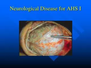
APD testing in 1 Reactive pupil • Upper series • Left. Pupils in dim light. • Middle. A normal response is seen when light shown in the OD • Right. When light is presented to OS the right pupil dilates. This is (+) for an APD. This indicates a probable optic nerve or retinal lesion. • Lower series • Left. Pupils in dim light. • Middle. A normal response is seen when light is shown in the OD • Right. When light is presented to OS the right pupil does not change size or re-dilate. There is no APD.
Pupils • Horner’s • Decrease in sympathetic innervation – miotic pupil • Adie’s Tonic pupil • Parasympathetic paralysis – dilated pupil • Pupillary Light-near dissociation • Argyll Robertson Pupil – miotic pupil • Parinaud’s Syndrome (Dorsal Midbrain) • Bilateral – Mid-dilated pupils • Light-near dissociation • Associated Supranuclear Upgaze palsy
Horner’s Syndrome • Disturbance in the sympathetic pathway • Central: Hypothalamus to the 1st synapse in Ciliospinal center of Budge, (C8-T2?) of the spinal cord • Cause - Stroke, MS, Vertebral artery dissection • Pre-ganglionic: Over the apex of the lung to 2nd synapse in the neck at the Superior cervical ganglion • Cause – Lung tumor, Neck SX (trauma), Breast tumor, thyroid adenoma • Post-ganglionic: from the neck through the cavernous sinus (near the carotid) to the muscle of Mueller • Carotid artery dissection, Tumor/Surgery of neck, cluster or migraine headaches, Cavernous sinus inflammation • Triad: miosis, ptosis, anhydrosis(post-ganglionic) • Miosis >> in dim illumination (doesn’t dilate normally)
Horner’s Syndrome • Cocaine test 2%-10%: • If no dilation/less than normal: (+) result • If dilation present: (-) result • Hydroxyamphetamine 1% (Paradrine): • Used to distinguish Central/Pre-ganglionic from Post-ganglionic lesion • Central/Pre-ganglionic – will dilate because NE is released from nerve endings • Post-ganglionic lesion – will NOT dilate because no NE is released • Only 40-97% sensitive is determining location Need to wait a minimum of 24-48 between performing the different tests
Horner’s Management • Work-up • Take a good history • Headaches, surgery/trauma, pain (neck), etc. • CBC with diff • CT scan of chest – to evaluate for apical lung tumor (Pancoat’s tumor) • MRI of brain & neck • Carotid doppler ultrasound vs MRA of head/neck
Adie’s Tonic Pupil • Dilated pupil > in bright lights (doesn’t constrict) – pt may be photophobic • Minimal or no Reaction to light • May become smaller over time • Slow, tonic constriction to convergence with (slow redilation) – blurred near vision • May notice vermiform (sectoral) movement of iris • More common in younger women>men • May have absence of deep tendon reflex (Adie’s Syndrome)
Adie’s Tonic Pupil • Benign condition, but need to r/o out other causes of light-near dissociation • Argyll-Robertson pupil (usually extremely miotic) • Parinaud’s Syndrome (Bilateral; convergence normal (not tonic), Paralysis of upgaze common • Etiology: idiopathic, viral, trauma, or diabetes mellitus • Pilocarpine supersensitivity 0.125% - will constrict a Adie pupil (will not constrict a normal pupil)
Argyll-Robertson Pupils • Bilateral • Small (often very small) • Poor/if any reaction to light • Normal convergence • Etiology: Neuro-syphilis (tertiary) • Exam- look for other signs (interstitial keratitis, salt-n-pepper fundus, papillitis, uveitis • Work-up: VDRL (RPR) & FTA-ABS (MHA-TP) & consider lumbar puncture to look for Spirochete in CSF
Parinaud’s Syndrome • AKA: Dorsal midbrain syndrome • Bilateral • Light-near dissociation • Normal (not tonic) convergence • Mid-dilated pupils • Inability to look up (supranuclear palsy) • Convergence retraction nystagmus • Pineal tumors, hydrocephalus, and MS are main causes • Need to obtain MRI/CT is suspected
Diplopia & Motility deficits • CN III palsy • CN IV palsy • CN VI palsy • Multiple Cranial nerve Palsies – Cavernous sinus syndrome
Diplopia • Questions to ask : • Does it go away when covering either eye • Monocular (Ref. Error, Corneal, Cataract, Retinal) • Binocular (Most often EOM &/or neurological) • Vertical vs. horizontal vs. oblique • Onset – gradual vs. rapid • Pain associated or not • Is the double vision slightly separated or extensive? • Small vs. Large angle • Is it worse when looking in other directions? • Comitant vs. Non-comitant
CN III Palsy • Innervates SR, IR, MR, IO, and levator • Superior division: SR & levator • Inferior division: IR, MR, IO,& also carries pupillary fibers to the iris sphincter • Can have external (motility impaired) or internal ophthalmoplegia (pupil affected) • Need to describe if complete or partial CN III palsy & if pupil involved or pupil-sparing
CN III Palsy • Diplopia will vary depending on involvement • Complete Palsy – eye is down & out • Superior division palsy – inability to look up with ptosis • Inferior division – inability to look adduct or look down (pupil may be involved) • Pupil-involved – fixed, dilated pupil • Pupil-sparing – normal reaction
CN III Palsy • Pupillary fibers run peripheral of the nerve • Less likely affected in microvascular etiology (DM,HTN, Hyperlipidemia) • More likely affected if compressive lesion or aneurysm pressing on the nerve & pupil fibers • Painful Oculomotor palsy involving the pupil is usually due to aneurysm (often PCOM) • Complete Oculomotor palsy that spares the pupil is usually caused by diabetic neuropathy.
CN III Palsy • Management: • Pupil involved – STAT MRI &/or MRA • Pupil sparing – if associated vascular disease, can monitor closely or may still want to get MRI • R/O out MG (with Tensilon test) • Need to r/o GCA, order ESR & CRP
CN IV palsy • Innervation of SO • Involved eye will be hypertrophic • Diplopia is oblique • May have head tilt to contralateral side of affected eye with small chin tuck • Only nerve to exit brainstem dorsally, so is highly susceptible to head trauma • Other etiologies: Vascular, infection, and aneurysm
CN IV palsy • Work-up • Isolate with Parks 3 step test or Maddox rod test • Ask about history of head trauma • R/O out MG & GCA (is suspected) • Check BP, blood sugar (if vascular suspected) • Consider MRI if younger patient with no history of head trauma or if it does NOT resolve within 3 months • Management: • If newly acquired: Follow-up q 1-3 months (based on etiology) • If small angle & stable – prisms can be used • Occlusion patch can be used to relieve symptoms (if no risk of amblyopia)
CN VI palsy • Innervates Lateral rectus • Longest intracranial course • Horizontal diplopia • Esotropia • Limited abduction • May have head turn to ipsilateral side • Etiology: Vascular**, trauma, inflammatory/infection, tumor, congenital
CN VI palsy • Work-up & Management: Same as CN IV palsy • Congenital CN IV palsy: • Duane’s (Type I): - • limited abduction (normal adduction), globe retraction on adduction, and narrowing palpebral fissure on eye movement • Mobius syndrome: • absence or underdevelopment of the 6th and 7th cranial nerve. So will lack facial expression and will have esotropia (often bilateral)
Multiple Ocular Nerve Palsies • Cavernous Sinus Syndrome • On either side of the pituitary gland • CN III, IV, and V are in the lateral wall • CN VI run through the body • Internal carotid runs centrally
Multiple Ocular Nerve Palsies • Cavernous Sinus (CS) Syndrome • Mass lesions (tumor or aneurysm) within the cavernous sinus often produces multiple CN involvement, pain (CN V), possible Horner’s syndrome • Etiology: Meningioma, Pituitary apoplexy, Metastatic lesions, Arteriovenous fistula, intracavernous aneurysm, mucomycosis (ie. Diabetics), cavernous sinus thrombosis • Management: STAT CT/MRI
Multiple Ocular Nerve Palsies • Superior Orbital Fissure Syndrome (Tolosa-Hunt Syndrome ) • Location is at the most anterior border of CS • Similar signs as Cavernous Sinus (CS) Syndrome, except much more painful unilateral ophthalmoplegia • Etiology: granulomatous infiltration of nerves and muscles • Orbital Apex Syndrome • Location is in orbital apex • Proptosis is common • Same CN involved but also optic nerve involvement common – must suspect if Optic neuropathy with multiple CN palsies
Intranuclear Ophthalmoplegia (INO) • Lesion at the MLF • Adduction deficit (on ipsilateral side) • Abduction nystagmus (in contralateral eye) • Often associated with what disease?
1 ½ syndrome • Lesion at the PPRF or the sixth nerve nucleus AND at the MLF • Ipsilateral gaze palsy with a contralateral INO • Ipsilateral eye can not adduct or abduct (no lateral movement) • Contralateral eye can ONLY Adduct • MLF damage results in – • No ipsilateral adduction (no MR innervation) • Abducens nucleus damage results in – • No ipsilateral abduction (no LR innervation) • No contralateral adduction (no MR innervation)
Myasthenia Gravis (MG) • Causes by antibodies interfering with acetylcholine receptors – not allowing nerve signals • Is a systemic condition • Symptoms/Signs: • Ptosis (worse at then end of the day) • Diplopia (worse at then end of the day) • Cogan’s lid twitch – (upward overshoot lid twitch when shifting from inferior to primary gaze) • Facial muscle weakness • Limb weakness • Difficulty swallowing or breathing*** (life threatening) • Change in voice
Myasthenia Gravis (MG) • Testing/Management: • Tensilon (endophonium) IV – will cause reversal of lid & motility disorders immediately • Ice test – Ice pack over ptotic lid for ~2 min will improve ptosis • Rest test (sleep test) – keep eyes close for 20 minutes or take small nap and often improves • Consult with PCP to have Ach receptor antibody essay test performed & systemic management
Common VF defects • Hemianopia or quadranopia • “PITS” • More congruous – closer to occipital lobe • Closer to occipital tip – closer to macula (more field involved) • Deeper in occipital lobe – more peripheral VF involvement • More incongruous – more anterior in visual pathway • Bitemporal defects • Chiasmal • Altitudinal • Monocular – ION • Bilateral – bilateral occipital lobe lesions