Download
1 / 41
430 likes | 572 Views
LOWER BACK PAIN. Erestain-Garan. CASE. Age: 45 years old CC: Lower back pain Occupation: office secretary. CHRONOLOGY OF EVENTS. NEUROLOGIC EXAMINATION FINDINGS. NEUROLOGIC EXAMINATION FINDINGS. Patient in left lateral decubitus with knee flexed

E N D
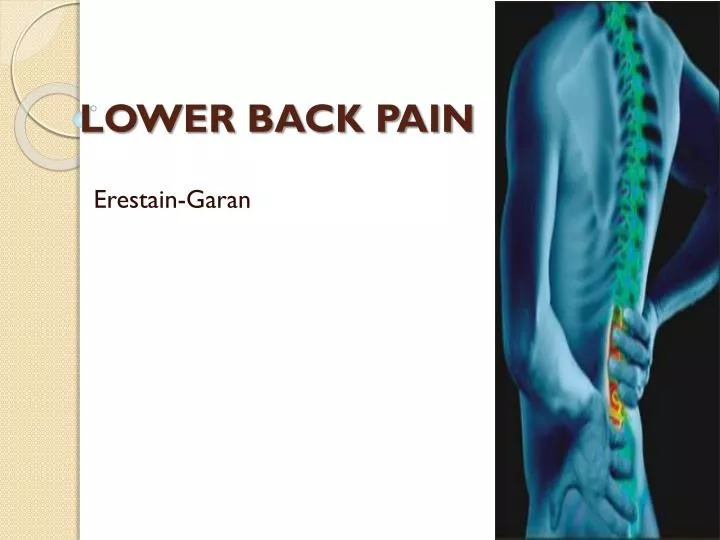
LOWER BACK PAIN Erestain-Garan
CASE • Age: 45 years old • CC: Lower back pain • Occupation: office secretary
NEUROLOGIC EXAMINATION FINDINGS • Patient in left lateral decubitus with knee flexed • Numbness: back of the right calf muscle, lateral heel, foot and toe • Weakness: right plantar flexion of foot and toes • Difficulty of walking on toes on the right • Atrophy: Right gastrocnemius and soleus muscles • Knee jerk: (++) both right and left • Ankle jerk: (++) left; (absent) right • Babinski: (-) both right and left • The rest of the neurological exam is within normal limit
SALIENT FEATURES • Age: 45 years old • sudden snap and pain in the LEFT LUMBAR AREA • Patient in left lateral decubitus with knee flexed • Numbness: back of the right calf muscle, lateral heel, foot and toe • Weakness: right plantar flexion of foot and toes • Difficulty of walking on toes on the right • Atrophy: Right gastrocnemius and soleus muscles • Knee jerk: (++) both right and left • Ankle jerk: (++) left; (absent) right • Babinski: (-) both right and left
Diagnosis of Spinal Injury • Spinal Injury is present if: • The person complains of severe pain in his or her neck or back. • An injury has exerted substantial force on the back or head. • The person complains of weakness, numbness or paralysis or lacks control of his or her limbs, bladder or bowel. • The neck or back is twisted or positioned oddly.
Spinal Cord Injury • Possibility of a SCI • Pain • Numbness • Difficulty with limb movements
Nature of the Problem • Non-traumatic Spinal Cord Injury • Age • Pain – left lumbar area • Resulted from normal physical strain (lifting a box of papers)
Lumbar Spine • Various forces that could be applied on the spine • nerve pathways narrowing and causing nerve impingement, inflammation, and pain
Key muscles = level of injury • C5 - Elbow flexors (biceps, brachialis) • C6 - Wrist extensors (extensor carpi radialis longus and brevis) • C7 - Elbow extensors (triceps) • C8 - Finger flexors (flexor digitorum profundus) to the middle finger • T1 - Small finger abductors (abductor digiti minimi) • L2 - Hip flexors (iliopsoas) • L3 - Knee extensors (quadriceps) • L4 - Ankle dorsiflexors (tibialis anterior) • L5 - Long toe extensors (extensors hallucis longus) • S1 - Ankle plantar flexors (gastrocnemius, soleus)
Numbness in the back of the R calf muscle (S1-S2) • Atrophy of R gastrocnemius and soleus Numbness in the lateral heel (S1) Numbness in the foot and toe (L4-L5, S1-S2)
Numbness in the foot and toe (L4-L5, S1-S2) Weakness of the R plantar flexion of foot and toes Difficulty walking on toes on the R
What is the difference between a radicular and myelopathic manifestations and what is the significance of each in relation to the signs and symptoms and clinical management?
Radiculopathy • Pain and numbness involving the degeneration or inflammation of the spinal nerve roots • usually without objective signs of neurologic dysfunction • Myelopathy • involves degeneration or any disease of the spinal cord
In Radiculopathy, compression of single root may not cause significant sensory loss (due to overlap of dermatomes in the body) • The main symptom: sharp, burning pain or “shooting pains”. • Follow a dermatomal distribution, accompanied by paresthesias, and loss of muscle power innervated by the root.
Symptoms of myelopathy would usually depend on the cause and severity of the condition • Trauma, herniated disc, OA of the spine, and tumors cause myelopathy • Symptoms: pain, loss of sensation or movement, decreased spinal range of motion, weakness, and deformity
Significance • From the signs and symptoms along with the PE results, we can achieve the correct diagnosis of our patient. • The persisting signs and symptoms would help us determine the appropriate mangement.
How does one localize the lesion based on anatomical diagnosis and other ancillary procedures?
Lower Motor Neuron Lesion • In the Case: • Patient has the following LMN symptoms: • (+) Atrophy of right gastrocnemius muscle and soleus muscles • (+) Areflexia in Right ankle jerk • (-) Babinski bilaterally
Lumbar Radiculopathy L5-S1 • In the patient: • Numbness in the back of the right calf muscle, lateral heel, foot and toe • Weakness of the right plantar flexion of foot and toes • Difficulty walking on toes on the right • Right ankle jerk absent
Numbness in back of the right calf muscle, lateral heel, foot and toe
Numbness in lateral heel, foot and toe Both knee jerks are (++) Right ankle jerk absent, left ankle jerk (++)
Diagnosis • If no improvement in symptoms have occurred in six weeks or red flags are present, imaging is appropriate. • CT scan used to evaluate the bony anatomy in the lumbar spine, which can show how much space is available for the nerve roots. • NEUROFORAMEN- vulnerable point of compression • MRI scan useful for determining where the nerve roots are being compressed ; shows the details of soft-tissue structures, like nerves and discs.
Diagnosis • MR neurography • modified MRI technique providing better pictures of the spinal nerves and the effect of compression on these nerves. • may help in diagnosis and treatment of sciatica/lumbar radiculopathy.
Treatment • goals of treatment : • relieve pain • prevent or reduce stress on the disc • maintain normal function • ranges from conservative therapies to surgical interventions
Conservative Treatment • Most treatment plans involve a combination of self-administered treatments, medications, and therapeutic measures. Self-administered treatments include the following: • Learn/practice proper posture and body mechanics • Rest and restrict activities • Limited bed rest to take pressure off the spine • Mild activity (exercise) such as walking, biking, and swimming • Apply cold and/or hot packs • Wear a brace for support (may not be helpful in all cases)
Conservative Treatment • Therapeutic treatments for DDD include the following: • Chiropractic treatment to manipulate the spine • Acupuncture to relieve pain • Massage therapy to relieve muscle spasms and tension • Physical therapy to improve function and increase flexibility and strength
Medications • Are used to supplement conservative therapy. • Non-steroidal anti-inflammatory drugs (NSAIDs; e.g., aspirin, ibuprofen, naproxen) • Pain relievers (e.g., acetaminophen) • Muscle relaxants • Spinal injections (anesthetics or corticosteroids) • Antidepressants • Sleep aids • Other non-surgical treatments • ultrasound therapy : uses sound waves to warm the area, increase blood flow, and relieve discomfort • transcutaneous electrical nerve stimulation (TENS): uses electrical stimulation of the nerve to interrupt pain signals
Surgical • Primary reasons for surgery are to: • relieve pressure on a nerve root or the spinal cord • stabilize an unstable or painful vertebral segment • prevent or limit radiculopathy (nerve damage) • reduce deformity or curvature of the spine (e.g., scoliosis)
Surgical • Discectomy and fusion • involves removing the damaged intervertebral disc and replacing it with a piece of bone or another material • this replacement fuses with the adjacent vertebrae • Corpectomy • a section of the vertebrae and discs is removed to create more space for the remainder of the spine • A bone graft and/or metal plate with screws – attached to stabilize the spine • Facetectomy, laminotomy, and spinal laminectomy • procedures that involve removing a portion of the bony structure of the spine to relieve pressure on the nerve roots • Foraminotomy and laminoplasty can be used to enlarge areas of the spinal column to make more room for the nerves and spinal cord
Surgical • Micro-discectomy • removes a disc through a very small incision using a microscope. • Percutaneous disc decompression • reduces or eliminates a small portion of the bulging disc through a needle inserted into the disc, minimally invasive • Spinal decompression • A non-invasive procedure that enlarges the Intra Vertebral Foramen (IVF) by aiding in the rehydration of the spinal discs. • Spinal laminectomy • relieves pressure of spinal stenosis • part of the lamina is removed or trimmed to widen the spinal canal and create more space for the spinal nerves.
Indications for Surgery Surgery may be recommended: • If the conservative treatment options do not provide relief within two to three months. • If leg or back pain limits normal activity • If there is weakness or numbness in the legs • If it is difficult to walk or stand, or if medication or physical therapy are ineffective, surgery may be necessary, most often spinal fusion.
Lumbar surgery • indicated in patients with severe spinal stenosis, in those with intractable pain, and in patients in whom an appropriate 6- to 12-month nonoperative course of treatment fails. • In elective cases, other conservative modalities should have been tried and observed to fail. • In cases of cervical disk disease with radiculopathy • indications for surgical treatment are intractable pain, progressive motor or sensory deficit, or symptoms refractory in a reasonable period of nonoperative therapy • In cases of cervical disk disease with myelopathy • early surgery to decompress the spinal cord is recommended to arrest progression if the clinical and radiographic changes are well correlated
References: • http://www.cedars-sinai.edu/515.html • http://www.cedars-sinai.edu/889.html • http://www.cedars-sinai.edu/5757.html • http://www.neurologychannel.com/degenerative-disc-disease/treatment.shtml • http://www.dcmsonline.org/jax-medicine/1999journals/april99/degenerative.htm • http://emedicine.medscape.com/article/1265453-treatment