Download
1 / 23
240 likes | 728 Views
Muscular Dystrophies. Noor Anam Al Salihi. Muscular Dystrophies. A group of hereditary progressive diseases each with unique phenotypic and genetic features. How to differentiate MYOPATHY from NEUROATHY? Myopathy Neuropathy

E N D
Muscular Dystrophies Noor Anam Al Salihi
MuscularDystrophies • A group of hereditary progressive diseases each with unique phenotypic and genetic features. • How to differentiate MYOPATHY from NEUROATHY? MyopathyNeuropathy -Weakness most marked proximally - Distally -No muscle wasting or depression of - There is tendon reflexes until at least advanced stage. -Normal abdominal and plantar reflexes - Abnormal -No sensory loss or sphincter disturbances. - May be present.
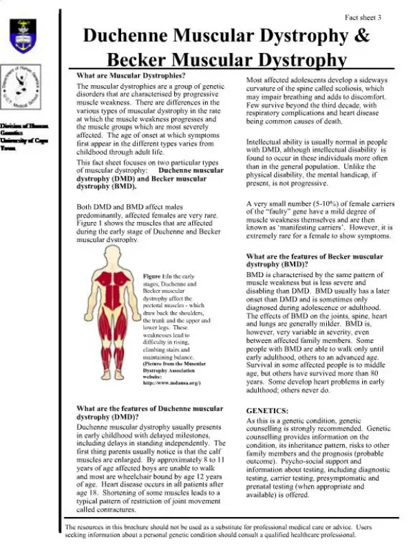
Duchenne • XR., sometimes called Pseudohypertrophic muscular dystrophy. • Incidence 30/100,000 live born males. • Clinical Features: • Duchenne dystrophy is present at birth, but the disorder usually becomes apparent between ages 3 and 5. • The boys fall frequently and have difficulty keeping up with friends when playing. Running, jumping, and hopping are invariably abnormal. • By age 5, muscle weakness is obvious by muscle testing. • On getting up from the floor, the patient uses his hands to climb up himself (Gowers' maneuver).
Gowers' sign showing a patient using arms to climb up the legs in attempting to get up from the floor.
Contractures of the heel cords and iliotibial bands become apparent by age 6, when toe walking is associated with a lordotic posture.
Loss of muscle strength is progressive, with predilection for proximal limb muscles and the neck flexors; leg involvement is more severe than arm involvement. • Between ages 8 and 10 walking may require the use of braces; joint contractures and limitations of hip flexion, knee, elbow, and wrist extension are made worse by prolonged sitting. By age 12, most patients are wheelchair dependent. • Contractures become fixed, and a progressive scoliosis often develops that may be associated with pain. The chest deformity with scoliosis impairs pulmonary function, which is already diminished by muscle weakness. By age 16 to 18, patients are predisposed to serious, sometimes fatal pulmonary infections. Other causes of death include aspiration of food and acute gastric dilation.
Cardiac cause of death is uncommon despite the presence of a cardiomyopathy in almost all patients. Congestive heart failure seldom occurs except with severe stress such as pneumonia. Cardiac arrhythmias are rare. • Intellectual impairment in Duchenne dystrophy is common; the average intelligence quotient (IQ) is ~1 SD below the mean. Impairment of intellectual function appears to be nonprogressive and affects verbal ability more than performance.
Lab. Features: • Serum CK levels are invariably elevated to between 20 and 100 times normal. The levels are abnormal at birth but decline late in the disease because of inactivity and loss of muscle mass. • EMG demonstrates features typical of myopathy. • The muscle biopsy shows muscle fibers of varying size as well as small groups of necrotic and regenerating fibers. Connective tissue and fat replace lost muscle fibers. • A definitive diagnosis of Duchenne dystrophy can be established on the basis of dystrophin deficiency in a biopsy of muscle tissue or mutation analysis on peripheral blood leukocytes.
Treatment • Glucocorticoids, administered as prednisone in a dose of 0.75 mg/kg per day, significantly slow progression of Duchenne dystrophy for up to 3 years. • Duchenne disease may benefit from novel therapies that either replace the defective gene or missing protein or implement downstream corrections (e.g., skipping mutated exons or reading through mutations that introduce stop codons).
Becker MuscularDystrophy • This less severe form of X-linked recessive muscular dystrophy results from allelic defects of the same gene responsible for Duchenne dystrophy. • Becker muscular dystrophy is ~10 times less frequent than Duchenne, with an incidence of about 3 per 100,000 live-born males.
Clinical Features • The pattern of muscle wasting in Becker muscular dystrophy closely resembles that seen in Duchenne. Proximal muscles, especially of the lower extremities, are prominently involved. As the disease progresses, weakness becomes more generalized. Significant facial muscle weakness is not a feature. Hypertrophy of muscles, particularly in the calves, is an early and prominent finding. • Most patients with Becker dystrophy first experience difficulties between ages 5 and 15 years, although onset in the third or fourth decade or even later can occur. By definition, patients with Becker dystrophy walk beyond age 15, while patients with Duchenne dystrophy are typically in a wheelchair by the age of 12. Patients with Becker dystrophy have a reduced life expectancy, but most survive into the fourth or fifth decade.
Mental retardation may occur in Becker dystrophy, but it is not as common as in Duchenne. Cardiac involvement occurs in Becker dystrophy and may result in heart failure; some patients manifest with only heart failure. Other less common presentations are asymptomatic hyper-CK-emia, myalgias without weakness, and myoglobinuria.
Laboratory Features • Serum CK levels, results of EMG, and muscle biopsy findings closely resemble those in Duchenne dystrophy. The diagnosis of Becker muscular dystrophy requires Western blot analysis of muscle biopsy samples demonstrating a reduced amount or abnormal size of dystrophin or mutation analysis of DNA from peripheral blood leukocytes. • Genetic testing reveals deletions or duplications of the dystrophin gene in 65% of patients with Becker dystrophy, approximately the same percentage as in Duchenne dystrophy.
Treatment • The use of glucocorticoids has not been adequately studied in Becker dystrophy.
Limb-Girdle Muscular Dystrophy • LGMD type 1AD, type2 AR • The syndrome of limb-girdle muscular dystrophy (LGMD) represents more than one disorder. Both males and females are affected. • Onset ranging from late in the first decade to the fourth decade. • typically manifest with progressive weakness of pelvic and shoulder girdle musculature. Respiratory insufficiency from weakness of the diaphragm may occur, as may cardiomyopathy.
Emery-Dreifuss Muscular Dystrophy • There are two genetically distinct forms of Emery-Dreifuss muscular dystrophy (EDMD). One is inherited as an X-linked disorder, while the other is autosomal dominant • Clinically the conditions are closely related..
Clinical Features • Prominent contractures can be recognized in early childhood and teenage years, often preceding muscle weakness. The contractures persist throughout the course of the disease and are present at the elbows, ankles, and neck. • Muscle weakness affects humeral and peroneal muscles at first and later spreads to a limb-girdle distribution. • The cardiomyopathy is potentially life threatening and may result in sudden death. A spectrum of atrial rhythm and conduction defects includes atrial fibrillation and paralysis and atrioventricular heart block.
Laboratory Features • Serum CK may be elevated two- to tenfold. • EMG is myopathic. • Muscle biopsy shows nonspecific dystrophic features. • Immunohistochemistry reveals absent emerin staining of myonuclei in X-lined EDMD. • ECGs demonstrate atrial and atrioventricular rhythm disturbances.
Treatment • Supportive care should be offered for neuromuscular disability, including ambulatory aids, if necessary. Stretching of contractures is difficult. Management of cardiomyopathy and arrhythmias (e.g., early use of a cardiac pacemaker) may be life saving.
Myotonia Congenita • Myotonia: is the delay of muscle relaxation after contraction leading to apparent muscle stiffness. There are two types of Myotonia Congenita • Thomsen’s Disease: - AD - Usually presents from birth but symptoms may not develop until early childhood. - There is generalized myotonia without weakness. Muscle stiffness is enhanced by cold and inactivity and relieved by exercise. - Muscle hypertrophy, sometimes pronounced.
Beker’s Disease: - AR. - Later onset. - There is slight weakness and atrophy of distal muscles. Treatment Qunine sulfate, Procainamide, mexilitene, and phenytoin.