Download
1 / 1
10 likes | 81 Views
1. A. B. 2. *. R. L. 3. *. R. L. *. B. A. E-figure 2. MRI of lower limbs in patient IV-1 of pedigree A

E N D
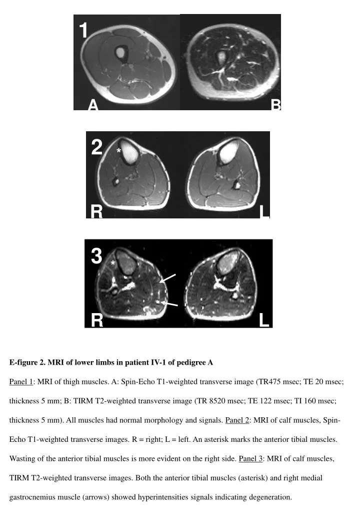
1 A B 2 * R L 3 * R L * B A E-figure 2. MRI of lower limbs in patient IV-1 of pedigree A Panel 1: MRI of thigh muscles. A: Spin-Echo T1-weighted transverse image (TR475 msec; TE 20 msec; thickness 5 mm; B: TIRM T2-weighted transverse image (TR 8520 msec; TE 122 msec; TI 160 msec; thickness 5 mm). All muscles had normal morphology and signals. Panel 2: MRI of calf muscles, Spin-Echo T1-weighted transverse images. R = right; L = left. An asterisk marks the anterior tibial muscles. Wasting of the anterior tibial muscles is more evident on the right side. Panel 3: MRI of calf muscles, TIRM T2-weighted transverse images. Both the anterior tibial muscles (asterisk) and right medial gastrocnemius muscle (arrows) showed hyperintensities signals indicating degeneration. A B *