Download
1 / 25
450 likes | 1.07k Views
Joint Hospital Surgical Grand Round United Christian Hospital Dr C Leung. Management of Barrett’s oEsophagus. Definition. A change in the normal squamous epithelium of the oesophagus to specialized intestinal metaplasia.

E N D
Joint Hospital Surgical Grand Round United Christian Hospital Dr C Leung Management of Barrett’s oEsophagus
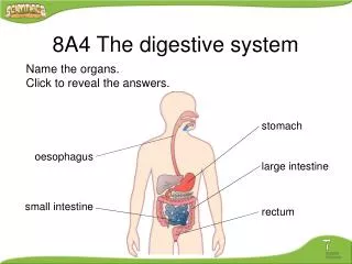
Definition • A change in the normal squamous epithelium of the oesophagus to specialized intestinal metaplasia Playford RJ. New British Society of Gastroenterology guidelines for the diagnosis and management of Barrett’s esophagus Gut 2006;55:442-3
Background • Prevalence • 1.6-5.6% • 10-15% in patients with reflux symptoms • Premalignant condition • 30-40fold increased risk of oesophagealCA
Etiology • Combined acid and bile reflux • > 50% of patients with GERD had abnormal levels of acid and bile in the oesophagus • Barrett’s esophagus patients have the highest level Fein M. Br J Surg 2006; 93: 1475-82
Risk of Adenocarcinoma • 0.25 to 0.4% per year • Nondysplasic : 3.86/1000 person years • Low-grade dysplaia: 7.66/1000 person years • High-grade dysplasia Occult carcinoma: 30%-40% of patients • 14.1/100 person years Sharma P. Clin Gastroenterol Hepatol 2006; 4: 566-72 Buttar NS. Gastroenterology 2001; 120: 1630-9
Endoscopic Evaluation • Prague classification • the maximal length (M) (including tongues) of Barrett esophagus • length of the circumferential Barrett segment (C) • For future endoscopic comparison Sharma P. Gastroenterology 2006; 131: 1392-9
Biopsies • Seattle protocol • 4 quadrant jumbo bx at 1cm intervals throughout whole length of Barrett’s • Separate target bx of any irregularities (nodules/erythema/ erosions) Reid BJ. Am J Gastroenterol 2000; 95: 3089-95.
Treatment rationale • Removal of diseased mucosa, not entire organ • Prevent disease progression to adenoCA
Treatment Options • Anti-reflux treatment -PPI -Fundoplication +/- surveillance • Endoscopic ablation • Photodynamic therapy (PDT) • Multipolar electrocoagulation • Argon Plasma Coagulation • Radiofrequency ablation (RFA) • Cryoablation resection • EMR/ ESD • Esophagectomy Symptomaticcontrol Cant reduce CA risk HGD / Tis , T1a adenoCA Multifocal, extensive HGD/ persistent HGD despite ablation/ ? CA
Acid Suppression with Surveillance • Acid suppression will noteliminate risk of adenocarcinoma/ consistent regression of Barrett’s ? Duration and dosage of PPI (indefinite) ?optimal frequency of surveillance
Anti-reflux Surgery • Fundoplication eliminates acid and bile reflux in > 90% of patients with Barrett’s oesophagus • Meta-analysis: 15.4% of patients undergone surgery will have regression of Barrett’s vs. 1.9% medically managed patients • Swedish Cohort study showed that RR of adenocarcinoma in patients undergone surgery was 14.1 vs. 6.3 for medical treatment Reduce risk of adenoCA ? Mixed evidences so far Oelschlager BK. Ann Surg 2003; 238: 458-64. Chang EY. Ann Surg 2007; 246: 11-21. Lagergren J. Gastroenterology 2010; 138: 1297-301
PPI vs fundoplication • Surgery can definitely treat reflux-related symptoms, but its role in protection against adenocarcinoma should be cautious • Effectiveness in eliminating reflux symptoms • Co- morbidities • Patient’s choice/ compliance • Medications S/E
Photodynamic Therapy • Injecting a light-sensitizing drug into patient, then expose the portion of oesophagus to a specific wavelength • Found NOTeffective in eliminating Barrett’s • ‘Buried glands’: a layer of normal-appearing squamous epithelium is present but under this layer, Barret’s metaplasia still present • Stricture • Phototoxicity Menon D. BMC Gastroenterol 2010; 10: 111.
Argon Plasma Coagulation • Systemic review: more effective than PDT, 3-month complete eradication 80% • Less complications like stricture or bleeding • Odynophagia 10% Li YM. Dig Dis Sci 2008; 53: 2837-46.
Radiofrequency Ablation • One of the best studied method • Applies bipolar electrical energy to mucosal surfaces, 10J for 1 second mucosa is ablated to submucosal level
Radiofrequency ablation • Need standardized FU as complete ablation with single treatment in only 70% of patients • FU OGD 3 months and 1 year, if not complete ablated repeat RFA
Radiofrequency ablation • Shaheen NJ (2009): Multicentre RCT • Can eliminate Barrett’s oesophagus with high grade dysplasia and reduce risk of oesophageal carcinoma • Wani S (2009): Meta-analysis • Reduction in carcinoma progression in high-grade dysplasia • Shaheen NJ (2011): Long term results • 3 years follow-up: complete eradication persist in 96% patients with high-grade dysplaia • Adenocarcinoma occurred in one per 181 patient-years of follow-up
Radiofrequency ablation • Promising results • S/E : esophageal stricture, GIB, chest pain • Sustaintially lower than those in photodynamic therapy • Long term data needed
Cryoablation • Endoscopically directed spray of liquid nitrogen at -196oC • Complete eradication of high grade dysplasia occurs in 68-97% of patients • Not well studied as RFA • ? Treat patient refractory to RFA Dumot JA. Gastrointest Endosc 2009; 70: 635-44. Shaheen NJ. Gastrointest Endosc 2010; 71: 680-5.
Endoscopic Mucosal Resection • when a visible nodule is present or only a short segment of Barrett’s is seen • substantial tissue for pathologist • treat Tis or T1a adenocarcinoma • Can combined with RFA With submucosal invasion, 20% risk of LN met If confined to mucosa ,<1% LN met
Endoscopic therapy • No single endotherapy achieve complete eradication without complications • Recurrence • For mucosal lesion • Buried metaplasia
Esophagectomy • ‘gold standard’ for high grade dysplasia and early adenocarinoma • 20-40% of patients harbour early adenocarcinoma in HGD (old data) • Mortality can be as low as 1% in high vol centre • Significant morbidity • For multifocal , too extensive HGD / intractable HGD /suspicious of carcinoma
Summary Barrett’s esophagus metaplasia LGD HGD Anti-reflux +surveillance OGD every3-5 year Anti-reflux+OGD every 6-12 months Repeat bx confirmed HGD Send to expert pathologist Endotherapy (ablative/EMR/ESD) If persist/ ? CA then esophagectomy
Take Home Messages • Barrett’s esophagus is a pre-malignant condition • Diagnosis relies on both endoscopic and histological findings • Management should be based on risks stratification • Emerging evidence on the use of endoscopic therapy • Treatment should be individualized