Download
1 / 73
920 likes | 1.38k Views
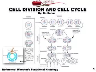
Cell division, cell growth, cell Cycle. MEIOSIS I: Separates homologous chromosomes. INTERPHASE. PROPHASE I. METAPHASE I. ANAPHASE I. Sister chromatids remain attached. Centromere (with kinetochore). Centrosomes (with centriole pairs). Chiasmata. Metaphase plate. Sister chromatids.

E N D
MEIOSIS I: Separates homologous chromosomes INTERPHASE PROPHASE I METAPHASE I ANAPHASE I Sister chromatids remain attached Centromere (with kinetochore) Centrosomes (with centriole pairs) Chiasmata Metaphase plate Sister chromatids Spindle Nuclear envelope Homologous chromosomes separate Microtubule attached to kinetochore Tetrad Chromatin Pairs of homologous chromosomes split up Chromosomes duplicate Tertads line up Homologous chromosomes (red and blue) pair and exchange segments; 2n = 6 in this example Figure 13.8 • Interphase and meiosis I 2. cross over 1. Synapsis (聯會) (synaptonemal complex)
MEIOSIS II: Separates sister chromatids TELOPHASE II AND CYTOKINESIS TELOPHASE I AND CYTOKINESIS METAPHASE II ANAPHASE II PROPHASE II Cleavage furrow Haploid daughter cells forming Sister chromatids separate Two haploid cells form; chromosomes are still double During another round of cell division, the sister chromatids finally separate; four haploid daughter cells result, containing single chromosomes Figure 13.8 • Telophase I, cytokinesis, and meiosis II
MITOSIS MEIOSIS Parent cell (before chromosome replication) Chiasma (site of crossing over) MEIOSIS I • A comparison of mitosis and meiosis Prophase I Prophase Chromosome replication Chromosome replication Tetrad formed by synapsis of homologous chromosomes Duplicated chromosome (two sister chromatids) 2n = 6 Tetrads positioned at the metaphase plate Chromosomes positioned at the metaphase plate Metaphase I Metaphase Sister chromatids separate during anaphase Homologues separate during anaphase I; sister chromatids remain together Anaphase Telophase Anaphase I Telophase I Haploid n = 3 Daughter cells of meiosis I 2n 2n MEIOSIS II Daughter cells of mitosis n n n n Daughter cells of meiosis II Sister chromatids separate during anaphase II
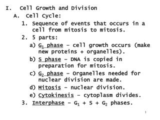
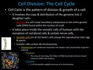
Cell cycle: • --- the life of a cell from the time it is first formed from a • dividing parent cell until its own division into two cells. • Smallest unit of life • all living things must reproduce • Cells replicate for growth, replacement, and repair • Cell division functions in reproduction, growth, and renewal. 200 µm 20 µm
Cell CycleThe Cell’s Time Clock Cytokinesis • Cell division requires Mitosis & Cytokinesis • Phases of a dividing cell’s life • interphase • cell grows • replicates chromosomes • produces new organelles & biomolecules • mitotic phase • cell separates & divides chromosomes • mitosis • cell divides cytoplasm & organelles • cytokinesis
Cell cycle • Cell has a “life cycle” cell is formed from a mitotic division cell grows & matures to divide again cell grows & matures to never divide again G1, S, G2, M liver cells G0 epithelial cells,blood cells, stem cells brain nerve cells
Interphase • Cell performs normal function • Three subphases: • G1: cell duplicates most organelles • S: quantity of DNA in the cell is doubled as chromosomes are replicated. Each chromosome has a pair of sister chromatids connected by a centromere that contains a kinetochore • G2: chemical components stockpiled • Nucleolus present
Nuclear division without a reduction in chromosome number Each new cell (daughter cell) will have the same quantity of DNA as the parental cell Why is this important? Mitotic events can be categorized into discrete stages based on what is happening to structure of the cell Stage include: Prophase Prometaphase Metaphase Anaphase Telophase Mitosis
Prophase(Including Prometaphase) • Pro • Three things visibly occur • Chromosomes condense (shorten) • Centrosomes migrate to the poles while producing spindle fibers • Nuclear membrane fragments
Metaphase Metaphase Plate • Meta • Chromosomes are moved by growing spindle fibers to the equator of the cell (metaphase plate) • Centrosomes are at the poles, nuclear membrane is gone
Anaphase • Ana • Centromere splits into two • Spindle fibers shorten from kinetochore end separating sister chromatids • Activated kinetochores “pull” chromatids along the spindle fibers and toward the poles
Telophase • Telo • Nuclear membrane reforms around each region of chromosomes • Nucleolus reforms • Cytokinesis (division of the cytoplasm) may occur
Vesiclesforming cell plate 1 µm Wall of patent cell Cleavage furrow Cell plate 100 µm New cell wall Contractile ring of microfilaments Daughter cells Daughter cells (a) Cleavage of an animal cell (SEM) (b) Cell plate formation in a plant cell (SEM) Cytokinesis divides the cytoplasm * Cleavage furrow * No cleavage furrow Actin + Myosin
Coordination of cell division • A multicellular organism needs to coordinate cell division across different tissues & organs • critical for normal growth, development & maintenance • coordinate timing of cell division • coordinate rates of cell division • not all cells can have the same cell cycle
Activation of cell division • How do cells know when to divide? • cell communication signals • chemical signals in cytoplasm give cue • signals usually mean proteins • activators • inhibitors experimental evidence: Can you explain this?
M anaphase metaphase telophase prophase C G2 interphase (G1, S, G2 phases) mitosis (M) cytokinesis (C) G1 S Frequency of cell division • Frequency of cell division varies by cell type • embryo • cell cycle < 20 minute • skin cells • divide frequently throughout life • 12-24 hours cycle • liver cells • retain ability to divide, but keep it in reserve • divide once every year or two • mature nerve cells & muscle cells • do not divide at all after maturity • permanently in G0
sister chromatids centromere single-stranded chromosomes double-stranded chromosomes There’s noturning back, now! Overview of Cell Cycle Control • Two irreversible points in cell cycle • replication of genetic material • separation of sister chromatids • Checkpoints • process is assessed & possibly halted
Cell Cycle Regulation • Cell cycle events are triggered by the cell-cycle control system; a set of molecules found in the cytoplasm affected by internal and external controls • Checkpoints in G1, G2, and M phases of the cycle • G1 checkpoint is most critical. May throw cells out of cyclic phase into G0, never to divide again
Other Internal and External Factors • Internal • M checkpoint does not proceed until signal is received that all kinetochores are attached to spindle microtubules • External • Growth factors: cycle will not proceed if requirements are not met • Social signals • Density-dependent inhibition: under crowded conditions chemical requirements are insufficient to allow cell growth • Anchorage dependence: some cells must be attached to a substrate in order to replicate • DNA damage inhibits growth
External signals: ex. Growth factors ~ Cells fail to divide if an essential nutrient is left out of the culture medium. ~ GFs trigger a signal transduction pathway that allows the cells to pass the G1 checkpoint and divide. PDGF PDGF receptor cell Signal transduction Cell division
External signals • Growth factors • coordination between cells • protein signals released by body cells that stimulate other cells to divide • density-dependent inhibition • crowded cells stop dividing • each cell binds a bit of growth factor • not enough activator left to trigger division in any one cell • anchorage dependence • to divide cells must be attached to a substrate • “touch sensor” receptors
Cells anchor to dish surface and divide (anchorage dependence). When cells have formed a complete single layer, they stop dividing (density-dependent inhibition). If some cells are scraped away, the remaining cells divide to fill the gap and then stop (density-dependent inhibition). 25 µm External signals: physical factor Density-dependent inhibition of cell division ~ Crowded cells stop dividing single layer
Most animal cells exhibit anchorage dependence • In which they must be attached to a substratum to divide Anchorage dependence • * Cancer cells: • ~ Exhibit neither density- • dependent inhibition nor • anchorage dependence Normal cell ~ single layer Cancer cells do not exhibit anchorage dependence or density-dependent inhibition. 25 µm 25 µm
Growth factor signals growth factor nuclear pore nuclear membrane P P cell division cell surface receptor Cdk E2F protein kinase cascade P chromosome P Rb P E2F Rb nucleus cytoplasm
Internal signalof a Growth Factor • Platelet Derived Growth Factor (PDGF) • made by platelets in blood clots • binding of PDGF to cell receptors stimulates cell division in fibroblast (connective tissue) • heal wounds Don’t forget to mentionerythropoietin!(EPO)
G1 checkpoint Control system S G1 G2 M M checkpoint G2 checkpoint The sequential events of the cell cycle are directed by a distinct cell cycle control system, a cyclically operating set of molecules in the cell that both triggers and coordinates key events in the cell cycle. ~ similar to a clock The cell cycle is regulated at certain checkpoints by both internal and external controls.
Checkpoint control system • Checkpoints • cell cycle controlled by STOP & GO chemical signals at critical points • signals indicate if key cellular processes have been completed correctly
Checkpoint control system • 3 major checkpoints: • G1/S • can DNA synthesis begin? • G2/M • has DNA synthesis been completed correctly? • commitment to mitosis • spindle checkpoint • are all chromosomes attached to spindle? • can sister chromatids separate correctly?
Spindle checkpoint G2 / M checkpoint Chromosomes attached at metaphase plate • Replication completed • DNA integrity Inactive Active Active Inactive Cdk / G2cyclin (MPF) M cytokinesis APC C mitosis G2 G1 S Cdk / G1cyclin Inactive MPF = Mitosis Promoting Factor APC = Anaphase Promoting Complex Active G1 / S checkpoint • Growth factors • Nutritional state of cell • Size of cell
G1/S checkpoint • G1/S checkpoint is most critical • primary decision point • “restriction point” • if cell receives “GO” signal, it divides • internal signals: cell growth (size), cell nutrition • external signals: “growth factors” • if cell does not receive signal, it exits cycle & switches to G0 phase • non-dividing, working state
G0 phase • G0 phase • non-dividing, differentiated state • most human cells in G0 phase • liver cells • in G0, but can be “called back” to cell cycle by external cues • nerve & muscle cells • highly specialized; arrested in G0 & can never divide
Cell Cycle Checkpoints • If cell size inadequate • G1 or G2 arrest • If nutrient supply inadequate • G1 arrest • If an essential external stimulus is lacking • G1 arrest (at R) • If the DNA is not replicated • S arrest • If DNA damage is detected • G1 or G2 arrest • If the spindle formation is improper, chromosome misalignment • M-phase arrest R
“Go-ahead” signals • Protein signals that promote cell growth & division • internal signals • “promoting factors” • external signals • “growth factors” • Primary mechanism of control • phosphorylation • kinase enzymes • either activates or inactivates cell signals
Cell cycle signals inactivated Cdk • Cell cycle controls • cyclins • regulatory proteins • levels cycle in the cell • Cdk’s • cyclin-dependent kinases • phosphorylates cellular proteins • activates or inactivates proteins • Cdk-cyclin complex • triggers passage through different stages of cell cycle activated Cdk
Types of Cyclins and Cdks • There are many types of cyclins, but the 4 main ones are: • Cyclin D (G1 cyclin) • Cyclin E (S-phase cyclin) • Cyclin A (S-phase and mitotic cyclin) • Cyclin B (mitotic cyclin) • These are the 3 main cdks • Cdk4 (G1 Cdk) • Cdk2 (S-phase Cdk) • Cdk1 (mitotic Cdk) • The complex of Cdk1 and cyclin B is called mitosis promoting factor (MPF)a.k.a maturation promoting factor
Rise and fall of cyclins Cyclin Concentration Mitosis
Cdks and cyclins Cyclin-dependent kinases (Cdks) are enzymes that are present in the cell cytoplasm at all times. However, they are inactive unless they are bound by a specific partner-protein called a cyclin to form a Cdk-cyclin complex The amount of cyclins in the cell changes – because they get degraded A Cdk-cyclin complex will push the cell cycle forward.
Figure 19-35 Phosphorylation and Dephosphorylation in the Activation of a Cdk-Cyclin Complex
MPF: M-phase Promoting Factor • MPF is composed of two key subunits: Cdc2 and Cyclin B. • Cdc2 is the protein that encoded by genes which are required for passage through START as well as for entry into mitosis. • Cyclin B is a regulatory subunit required for catalytic activity of the Cdc2 protein kinase.
What does MPF do? The complex of Cdk1 and cyclin B is called mitosis promoting factor (MPF)
MPF activity is dependent upon Cyclin B • The cyclins were identified as proteins that accumulate throughout interphase and are rapidly degraded toward the end of mitosis. • It is suggested that they might function to induce mitosis, with their periodic accumulation and destruction controlling entry and exit from M phase.
MPF activity is dependent upon Cyclin B • Accumulation and degradation of cyclins
Figure 19-34 Fluctuating Levels of Mitotic Cyclin and MPF During the Cell Cycle