Download
1 / 110
1.1k likes | 1.4k Views
1. Which layer of the heart allows it to act as a pump ? a. Epicardium b. Myocardium c. Pericardium d. Endocardium. 2. Which slinglike structure supports the heart ? a.Pericardium b.Chordae tendineae c.Myocardium d.Endocardium. 3.

E N D
1 • Which layer of the heart allows it to act as a pump? • a. Epicardium • b. Myocardium • c. Pericardium • d. Endocardium
2 • Which slinglike structure supports the heart? • a.Pericardium • b.Chordaetendineae • c.Myocardium • d.Endocardium
3 • What is the hardest working cardiac chamber and therefore has the thickest myocardium? • a.Rightatrium • b.Rightventricle • c.Leftatrium • d.Leftventricle
4 • The right ventricle pumps blood to the • a. right atrium. • b. pulmonary veins. • c. pulmonary artery. • d. aorta.
5 • Vessel(s) that carry(ies) blood from the pulmonary capillaries to the left atrium is (are) the • a. Aorta • b. Pulmonary artery • c. Pulmonary veins • d. Vena cava
6 • The aorta receives blood from the • a. right ventricle. • b. pulmonary veins. • c. pulmonary artery. • d. left ventricle.
7 • Blood flows from the right atrium through which atrioventricular valve to the right ventricle? • a. Bicuspid • b. Mitral • c. Pulmonic • d. Tricuspid
8 • Which of the following structures “sees” unoxygenated blood? • a. Aorta • b. Left ventricle • c. Pulmonary artery • d. Pulmonary veins
9 • Which of the following “sees” oxygenated blood? • a. Vena cava • b. Pulmonary veins • c. Right atrium • d. Pulmonicvalve
10 • Chordaetendineae are not associated with which valve? • a. Aortic • b. Mitral • c. Tricuspid • d. Bicuspid
11 • With which of the following is “lubb-dupp” associated? • a. Myocardial contraction • b. Ventricular depolarization • c. Closing of heart valves • d. Diffusion of O2 from the lungs to the blood in the pulmonary capillaries
12 • Which of the following supplies oxygenated blood to the heart muscle? • a. Coronary arteries • b. Pulmonary artery • c. Pulmonary veins • d. Cardiac veins
13 • Where does the cardiac action potential (cardiac impulse) normally originate? • a. AV node • b. Purkinje fibers • c. Ectopic focus • d. SA node
14 • The pacemaker of the heart is located in the upper wall of the • a. right atrium. • b. right ventricle. • c. left atrium. • d. left ventricle.
15 • Referring to the ECG, the P wave represents • a. atrialcontraction. • b. ventricular relaxation. • c. atrialdepolarization. • d. atrialrepolarization.
16 • Referring to the ECG, the QRS complex represents ventricular • a. contraction. • b. repolarization. • c. relaxation. • d. depolarization.
17 • Which structure connects the cusps of the AV valves to the ventricles? • a. Purkinje fibers • b. AV node • c. Bundle of His • d. Chordaetendineae
18 • What are the conducting fibers that rapidly spread the electrical signal throughout the ventricles? • a. Bundle of His • b. Purkinje fibers • c. SA node • d. AV node
19 • Which of the following is least related to the mitral valve? • a. Left heart • b. Bicuspid • c. Semilunar • d. Chordaetendineae
20 • All of the following are electrical terms except • a. sarcomere. • b. depolarization. • c. action potential. • d. repolarization.
21 • Which of the following must precede ventricular contraction? • a. Ventricular relaxation • b. “Lubb-dupp” • c. Ventricular depolarization • d. Closing of the AV valves
22 • Which of the following semilunar valves “sees” oxygenated blood? • a. Mitral • b. Bicuspid • c. Aortic • d. Pulmonic
23 • The correct sequence is: blood flows from the right atrium to the right ventricle to the pulmonary artery to the • a. pulmonary veins. • b. coronary arteries. • c. pulmonary capillaries. • d. vena cava.
24 • What event causes the pulmonic valve to open? • a. The P wave • b. An increase in the pressure within the right ventricle • c. “Lubb-dupp” • d. Contraction of the chordaetendineae
25 • Which layer of the heart has actin, myosin, and intercalated discs? • a. Myocardium • b. Endocardium • c. Epicardium • d. Pericardium
26 • The pericardium is a part of the • a. myocardium. • b. diaphragm. • c. epicardium. • d. endocardium.
27 • The mitral and the bicuspid valves • a. are semilunar valves. • b. are both located on the right side of the heart. • c. “see” only unoxygenated blood. • d. are the same valves.
28 • The pulmonic and aortic valves • a. are atrioventricular valves. • b. “see” only oxygenated blood. • c. are attached to the ventricular walls by chordaetendineae. • d. are semilunar valves.
29 • The correct sequence is: blood flows from the pulmonary capillaries to the pulmonary veins to the left atrium to the left ventricle to the • a. vena cava. • b. pulmonary artery. • c. aorta. • d. circle of Willis.
30 • The atrioventricular node (AV node) • a. is the pacemaker of the heart. • b. is located in the upper part of the right atrium. • c. has a rate that is normally faster than the SA node. • d. delays the electrical signal coming from the atria into the ventricles.
31 • The purpose of which structure is to delay the spread of the signal from the atrium to the ventricles? • a. SA node • b. Purkinje fibers • c. Bundle of His • d. AV node
32 • Which of the following is an electrical event? • a. “Lubb-dupp” • b. Actin and myosin interaction • c. Murmur • d. Depolarization
33 • Which of the following is a result of ventricular contraction? • a. The AV valves open. • b. The semilunar valves close. • c. Blood is pumped into the pulmonary artery and the aorta. • d. Blood flows back into the atria.
34 • Under what condition is blood most likely to flow “backward” (e.g., from the left ventricle back into the left atrium)? • a. Pulmonary artery hypertension • b. Left ventricular hypertrophy • c. An insufficient mitral valve • d. Pulmonary edema
35 • What are ventricles “doing” during atrial contraction? • a. Pumping blood into the great vessels • b. Closing their AV valves • c. Relaxing • d. Opening their semilunar valves
36 • The Purkinje fibers • a. open valves. • b. pull on the cusps of the valves. • c. conduct electrical signals throughout the ventricles. • d. close valves.
37 • Which cavity first receives unoxygenated blood from the vena cava? • a. Right ventricle • b. Left atrium • c. Left ventricle • d. Right atrium
38 • What is the name of the valve that prevents backflow of blood • a. Mitral • b. Pulmonic • c. Semilunar • d. Tricuspid
39 • What is the cause of the heart sounds “lubb-dupp”? • a. Closing of the heart valves • b. Flow of blood through the coronary arteries • c. The electrical signal as it moves through the AV node • d. The firing of the autonomic nerves to the SA node
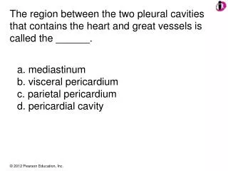
40 • Which of the following is not true of the heart? • a. The heart is located within the mediastinum. • b. The apex is located left of the sternal midline at the level of the fifth intercostal space. • c. The base of the heart is located at the level of the second rib. • d. The pericardium is composed of actin and myosin.
41 • Which of the following is least descriptive of the myocardium? • a. Cardiac muscle composed of actin and myosin arranged in sarcomeres • b. Thicker in the ventricles than the atria • c. Thicker in the left ventricle than the right ventricle • d. Thicker in the left atrium than the right ventricle
42 • Which of the following is a function of a valve? • a. Regulates the direction of the flow of blood through the heart • b. Regulates the amount of oxygen bound to hemoglobin • c. Regulates heart rate • d. Directs the movement of the cardiac impulse
43 • Which of the following is true of the structures of the electrical conduction system? • a. The AV valve is the pacemaker. • b. In normal sinus rhythm, the electrical signal arises within the SA node. • c. The His-Purkinje system spreads the electrical system from the right atrium to the left atrium. • d. The purpose of the AV node is to increase the speed at which the cardiac impulse moves from the atria to the ventricles.
44 • Which of the following is least true of the aortic valve? • a. It is also called the left semilunar valve. • b. It “sees” oxygenated blood. • c. Blood flows from the ventricle through this valve into the pulmonary artery. • d. An incompetent aortic valve allows blood to leak from the aorta back into the left ventricle.
45 • An accumulation of excess fluid in the pericardial space • a. causes external compression of the heart. • b. depresses the SA node, thereby eliminating pacemaker activity. • c. causes valvularstenosis. • d. causes a left-to-right shunt.
46 • A hole in the interventricular septum causes • a. a right-to-left shunt. • b. extreme cyanosis. • c. blood to shunt from the left ventricle to the right ventricle. • d. blood to shunt from the left ventricle to the pulmonary artery.
47 • Which structure “sees” oxygenated blood? • a. Tricuspid valve • b. Pulmonary artery • c. Pulmonary veins • d. Right semilunar valve
48 • Which group is incorrect? • a. Semilunarvalves: pulmonic, aortic • b. Atrioventricularvalves: tricuspid, bicuspid, mitral • c. Structures that carry oxygenated blood: pulmonary veins, left ventricle, aorta • d. Structures that carry unoxygenated blood: right ventricle, venaecavae, pulmonary veins
49 • Which group is incorrect? • a. Semilunarvalves: pulmonic, aortic • b. Structures that carry oxygenated blood: pulmonary veins, left ventricle, aorta • c. Layers of the heart: epicardium, myocardium, endocardium • d. Abnormal heart rates: tachycardia, bradycardia, normal sinus rhythm
50 • Which group is incorrect? • a. Atrioventricularvalves: tricuspid, bicuspid, mitral • b. Layers of the heart: epicardium, myocardium, endocardium • c. Abnormal heart rates: tachycardia, bradycardia • d. Parts of the conduction system: SA node, AV node, bundle of His, Purkinje fibers, medulla oblongata