Download
1 / 46
460 likes | 661 Views
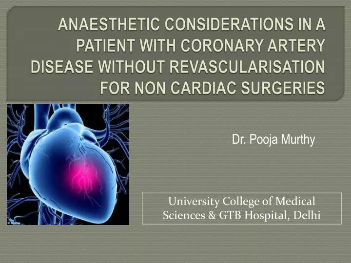
ANAESTHETIC CONSIDERATIONS IN A PATIENT WITH CORONARY ARTERY DISEASE WITHOUT REVASCULARISATION FOR NON CARDIAC SURGERIES. Dr. Pooja Murthy. University College of Medical Sciences & GTB Hospital, Delhi. INTRODUCTION. WHY THIS TOPIC ?

E N D
ANAESTHETIC CONSIDERATIONS IN A PATIENT WITH CORONARY ARTERY DISEASE WITHOUT REVASCULARISATION FOR NON CARDIAC SURGERIES Dr. Pooja Murthy University College of Medical Sciences & GTB Hospital, Delhi
INTRODUCTION WHY THIS TOPIC ? IHD is present in 30% of patients undergoing noncardiac surgeries. Increased perioperative cardiovascular mortality and morbidity. Increased in total health care expenditure. Increased chances of poor outcome in 1-2 yrs following surgeries
PERIOPERATIVE COMPLICATIONS OF IHD MOST IMPORTANT – Perioperative MI Incidence - <1% in patients who do not have CAD - 5 -15% in patients undergoing high risk surgeries Most perioperative MI occur in the first 24 – 48 hrs of surgery. Most common is NSTEMI. ( preceded by tachycardia and ST depression )
OTHER COMPLICATIONS These generally follow peri op MI. 1) Cardiac dysrhythmias: Ventricular fibrillation Ventricular tachycardia Atrial fibrillation Bradydysrhythmias and heart block
2) Pericarditis 3) Mitral regurgitation 4) Ventricular septal rupture 5) Heart failure and cardiogenic shock 6) Myocardial rupture 7) CVA
Pathogenesis IHD mainly occurs because of narrowing of the lumen of coronary arteries due to : Atherosclerosis (most common) Emboli Spasm Aortitis Cong. Abnormalities of coronary arteries
ABOUT IHD…. Coronary circulation normally supplies sufficient blood flow to meet the O2 demands of the myocardium in response to widely varying workloads. myocardial O2 supply (coronary blood flow myocardial O2 demand & O2 content) IMBALANCE IHD
PATHOPHYSIOLOGY Determinants of myocardial O2 demand are: MAJOR DETERMINANTS MINOR DETERMINANTS Myocardial contractility Heart rate Muscle shortening Wall stress Activation Determinants of myocardial Oxygen supply: Coronary blood flow Diastole
Determinants of myocardial oxygen supply/demand ratio Contractility % increase in MVO2 above resting value Heart rate Wall tension Muscle shortening Activation Basal metabolic requirement % increase of each factor above resting value
Classification of CAD Unstable Angina STEMI NSTEMI CAD Stable Angina Acute Coronary Syndromes
Manifestations of CAD… ISCHEMIC TYPE OF CHEST PAIN Partial occlusion / chronic narrowing of COR.ARTERY Complete occlusion of COR.ARTERY CSA ACS NO ST elevation 12 lead ECG Tp/CKMB - ve ST elevation Tp/CKMB + ve Tp/CKMB + ve UA Myocardial Infarction STEMI NSTEMI NEW ONSET / CHANGE FROM BASELINE
Evaluation and management of a patient with CAD for non cardiac surgery. Pre operative evaluation Intraoperative management Post operative management
Preoperative evaluation Goals: To know if the patient is on his/ her optimal medical regimen To know the severity of IHD To know the ischemic threshold Ventricular function
Medications Drug Interactions during Anaesthesia Nitrates Venodilation - hypotension Beta blockers Bradycardia with opioids Statins Liver dysfunction Antiplatelets Increased bleeding ACE I or ARBs Hypotension CCBs AV block with ß blockers Digitalis Toxicity Diuretics Hypokalemia (dysrhythmias)
Medications All cardiac medications (except ARBs) like beta blockers, calcium channel blockers, nitrates ,statins should be continued until the morning of surgery. Antiplatelets: aspirin or ADP antagonists which should be stopped at various time intervals (depending on the cardiac risk and risk of surgery )
Active cardiac conditions Unstable coronary syndromes - unstable/severe angina( CCS class III & IV) - recent MI( acute MI < 7 days,recent –7days to 1mth) Decompensated heart failure (NYHA class IV, new onset/worsening HF ) Significant arrhythmias (High degree heart blocks, ventricular arrhythmias symptomatic tachy and bradycardias ) Severe valvular disease (severe AS or symptomatic MS)
Clinical risk factors (CRIs) Detected of clinical risk factors as given by RCRI ( previously intermediate risk factors ). These include : h/o IHD h/o compensated / prior HF h/o CVD Renal insufficiency Diabetes mellitus
Surgery specific issues Surgical procedures : - emergency surgery has 2- 5% higher cardiovascular risk when compared to elective surgeries.
Cardiac risk stratification ( combined incidence of cardiac death and nonfatal MI )
Preoperative testing Preoperative Resting 12-Lead ECG CLASS I 1. Preoperative resting 12-lead ECG is recommended for patients with at least 1 clinical risk factor who are undergoing vascular surgical procedures. (Level of Evidence: B) 2. Preoperative resting 12-lead ECG is recommended for patients with known CHD, peripheral arterial disease, or cerebrovascular disease who are undergoing intermediate-risk surgical procedures. ( Level of Evidence: C)
Contd . . . Class II: Pre operative 12 lead ECG is reasonable in patients with no clinical risk factors who are undergoing vascular surgical procedures Pre operative 12 lead ECG is reasonable in patients with at least 1 clinical risk factor who are undergoing intermediate risk surgeries Presence of LV hypertrophy or ST segment depression on 12 lead ECG predicts adverse perioperative cardiac events.
Assessment of LV function : ( by ECHO, radionuclide angiography, contrast ventriculography ) No class I recommendation. It is reasonable( class II, C) in: Pts with dyspnoea of unknown origin Current or prior heart failure with worsening heart failure or change in clinical status.
Stress testing This includes: • Exercise stress testing – exercise ECG testing. • Pharmacological stress testing. • Nuclear myocardial perfusion imaging • Dobutamine stress echocardiography.
If a test is indicated…. Which test ? In ambulatory pts. – exercise ECG testing ( except in pts with abdominal aortic aneurysms) In pts with abnormal resting ECG( LBBB, LV hypertrophy with strain pattern, digitalis effect) – nuclear myocardial perfusion imaging. In pts who cannot exercise – non exercise stress test.
Intraoperative management Goals : • Prevent ischemia • Monitor for myocardial injury • Treatment of ischemia / infarction Premedication • Benzodiazepines • α2 agonists
Monitoring Routine monitors: 1. ECG – lead II, V5 ( 2 lead ) - lead II, V4, V5 ( 3 lead ) - lead II, V3 ,V5 ( 3 lead ) 2. Blood pressure 3. Pulse oximetry 4. Capnography 5. Temperature monitoring
Special monitors: Pulmonary artery catheter Trans esophageal ECHO Indicated in pts with hemodynamic instability..
INTRAOPERATIVE MANAGEMENT GOAL: To maintain a balance between myocardial oxygen demand and supply. Strategies : Optimize hemodynamics Correct Anaemia Maintain temperature (≥35 degree) Maintain blood glucose ≤150 mg/dl (Class IIa) Prevent pain
Intraoperative events that influencemyocardial O2 delivery and O2 requirement Decreased O2 delivery: Decreased coronary blood flow Tachycardia Diastolic hypotension Hypocapnia Coronary artery spasm Anemia Arterial hypoxemia Shift of O2 dissociation curve to left
Increased O2 requirement: Sympathetic stimulation Tachycardia Hypertension Increased myocardial contractility Increased afterload Increased preload
It is recommended to keep Heart rate and Blood pressure within 20% of awake value intraoperatively…
Intraop management contd… Anaesthesia technique: General anaesthesia, regional anaesthesia, monitored anaesthesia care – any technique can be used but hemodynamic stability must be ensured.
Induction of anaesthesia - Intravenous cannulation (pain free) - Premedication – opioids ( morphine, fentanyl ) , benzodiazepines - Inducing agents : Any intravenous inducing agents can be used except ketamine.
contd… Muscle relaxants: NDMRs / Succinylcholine can be used. NDMRs: Vecuronium , rocuronium, cisatracurium preferred – minimum/ no effect on HR and BP Atracuium – less preferred because of histamine release. Pancuronium - increases HR and BP and incidences of myocardial ischemia has been reported.
Contd… Prevention of intubation response: • Short duration of laryngoscopy ( < 15sec) • Laryngotracheal lidocaine • I V Lidocaine 1.5 – 2mg/kg – 2 to 3 min before intubation • I V Esmolol 0.5 – 1mg/kg – 90 sec before intubation • I VRemifentanil (1.0 g/kg) 1 min, Alfentanil (10–20 g/kg) 2–3 min, or Fentanyl (0.5–1.0 g/kg) 4–5 min • Diltiazem, oral clonidine(0.2mg), dexmeditomedine infusion (1µg/kg).
MAINTENANCE OF ANAESTHESIA • Volatile anaesthetic agents ( halothane less preferred because of myocardial depression ) • Maintenance of hemodynamic stability • Maintenance of euglycemia • Maintenance of normothermia • Adequate analgesia
Reversal of NMB : Anticholinesterase/ anticholinergic drug can safely be used Prevent extubation response Glycopyrrolate preferred over atropine.
Intra op management of myocardial ischemia Diagnosis : ST segment elevation / depression of at least 1mm on ECG Steps in management : Prompt and aggressive treatment of changes in heart rate and blood pressure is indicated
Increase in heart rate – beta blockers e.g. esmolol • Increase in BP – NTG • Decrease in BP – fluid infusion + sympathomimetic drugs • Unstable hemodynamic situation : • Circulatory support • Inotropes - Intra aortic balloon pump counter pulsation Plan early post op cardiac catheterization.
Post op management • Goal: - Maintain myocardial perfusion - Minimize haemodynamic stress • Things to be cautious about: • Intra op hypothermia – predisposes to post op shivering - pain, hypoxia, hypercarbia, sepsis, blood loss – increases myocardial O2 demand
Post op care: • Continue beta blockers and other medications • Prevention of hypovolemia and hypotension • Correction of anemia • Continuous ECG monitoring to detect silent ischemia
References ACC/ AHA 2007 Guidelines on perioperative cardiovascular evaluation and care for non cardiac surgeries. Kaplan’s textbook of cardiac anaesthesia – 5th edition Stoelting’s Anaesthesia and Coexisting disease – 5th edition Braunwald’s Heart Disease – 8th edition Miller’s Anaesthesia – 7th edition