Download
1 / 11
140 likes | 932 Views
Types of Microscopes . Contents Compound Microscope Dissecting Microscope Transmission Electron Microscope Scanning Electron Microscope Bibliography. Compound Microscope. More on Compound Microscopes. Compound Microscope.

E N D
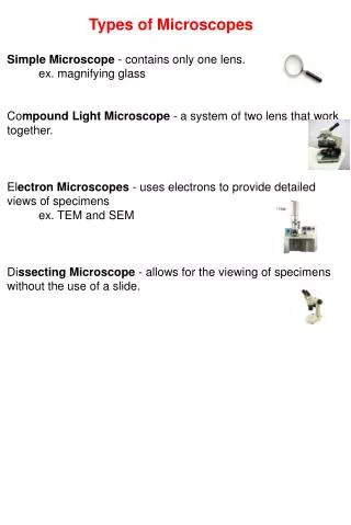
Types of Microscopes Contents Compound Microscope Dissecting Microscope Transmission Electron Microscope Scanning Electron Microscope Bibliography
Compound Microscope More on Compound Microscopes
Compound Microscope Hans Lippershey and his son, ZacchariasHanssen were experimenting with a variety of lenses. In the late 1590’s, they used many lenses in a tube and were amazed to see that the object at the end of the tube was magnified well beyond that of a magnifying glass. Little did they know, they had just invented the compound microscope. A Compound Microscope can be used for a number of different things, from finding medical miracles to a day at the park. Compound Microscope consists of an eyepiece which is usually 10x magnified followed by 10x, 40x and 100x objective lenses. A light then passes through the object to the objective lens, making an enlarged image of the object allowing you to see what the naked eye alone can’t.
Dissecting Microscope More on Dissecting Microscope
Dissecting Microscopes Philip O. Gravelle, a chemist, developed the Dissecting Microscope to compare bullets for identification in forensic science. It's history dates back to the 1920s. A Dissecting Microscope is a pair of microscopes placed next to each other, the optical paths of each microscope are connected together by the optical bridge, this helps the forensic examiners to simultaneously compare twothings.
Transmission Electron Microscope (TEM) More on Transmission Electron Microscopes
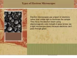
Transmission Electron Microscopes (TEM) The first TEM was built by Max Knoll and Ernst Ruska in 1931. TEM forms a majormethod in a range of scientific fields, in both physical and biological sciences. TEMsfind most use in cancer research, virology, materials science as well as pollution and semiconductor research. A TEM image of the polio virus.
Scanning Electron Microscope More on Scanning Electron Microscopes
Scanning Electron Microscope (SEM) Charles Oatley in 1952 created the Scanning Electron Microscope, but Max Knoll created the first prototype of the SEM in 1935. SEM is a type of electron microscope that images the objects surface by scanning it with a high-energy beam of electrons. pollen grains taken on an SEM
Bibliography Information/Images for Compound Microscopes: http://www.labessentials.com/Microscopes_Compound_Basics.htm http://leavingbio.net/Cell%20Structure_files/Cell%20Structure_files/image002.jpg Information/images for Dissecting Microscope http://dissectingmicroscopes.biz/dissecting-microscopes/the-dissecting-microscope-and-forensics/ http://www.clt.astate.edu/mhuss/stereoparts.jpg Information/Images for Transmission Electron Microscope http://www.udel.edu/biology/Wags/histopage/illuspage/lec1/iintro9.gif http://en.wikipedia.org/wiki/Transmission_electron_microscope Information/Images for Scanning Electron Microscope http://en.wikipedia.org/wiki/Scanning_Electron_Microscope http://www.purdue.edu/REM/rs/graphics/sem2.gif