Download
1 / 73
740 likes | 778 Views
Cardiomyopathy. Dr. Jamal Dabbas Interventional cardiologist & internist. Definition:. Group of diseases that primarily affect the heart muscle and are not the result of congenital, acquired, valvular, hypertensive , coronary arterial or pericardial abnormalities. Classification:

E N D
Cardiomyopathy Dr. Jamal Dabbas Interventional cardiologist & internist
Definition: Group of diseases that primarily affect the heart muscle and are not the result of congenital, acquired, valvular, hypertensive , coronary arterial or pericardial abnormalities
Classification: Dilated primary myocardial involvement Restrictive secondary myocardial involvement: Hypertrophic ( infective, Metabolic, Connective tissue disorders Infiltration, toxin, peripartum)
Cardiomyopathy • WHO Classification • anatomy & physiology of the LV • Dilated • Enlarged • Systolic dysfunction • Hypertrophic • Thickened • Diastolic dysfunction • Restrictive • Diastolic dysfunction • Arrhythmogenic RV dysplasia • Fibrofatty replacement
Cardiomyopathies are diseases of the muscle tissue of the heart. There are four major morphological types: dilated, hypertrophic, restrictive, and arrhythmogenic right ventricular cardiomyopathy.
In dilated cardiomyopathy, the most common type, ventricular output decreases eventually leading to failure of the left and right heart. The dilation can be seen on echocardiography, the most important diagnostic tool for all cardiomyopathies. Treatment is mostly similar to that of heart failure from other causes.
Hypertrophic cardiomyopathy can occur with or without left ventricular outflow obstruction. It is often asymptomatic, although arrhythmias and even sudden cardiac death can occur.
The obstructive type is also notable for signs of reduced blood flow (dyspnea, vertigo, syncope). It is important that patients avoid strenuous exercise. Symptomatic patients should be treated with beta blockers.
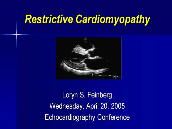
Restrictive cardiomyopathy is caused by the proliferation of connective tissue with subsequent atrial enlargement (but normal ventricles). Like dilated myopathy, restrictive cardiomyopathy produces left and right heart failure. Ejection fraction is usually normal, but diastolic filling is reduced on echocardiography.
Arrhythmogenic right ventricular cardiomyopathy affects primarily the right ventricle and is characterized by cellular breakdown and subsequent ventricular dilation. Symptoms are very variable although the hallmark finding is arrhythmia. There is no general best treatment as it depends entirely on individual factors such as the extent of the disease. All patients should avoid strenuous exercise.
Dilatedcardiomyopathy Definition: Dilatedcardiomyopathy(DCM)ischaracterizedbyleftventriculardilatationandsystolicdysfunctionintheabsenceofhypertension,coronaryarterydisease,valvedisease,congenitalheartdisease. Atrialdilationaswellasrightventriculardilationanddysfunctioncanalsodevelop.
DCM-IncidenceandPrognosis • Most common cardiomyopathy • Completerecoveryis rare • 50%diewithin2yrsand25%survive longerthan 5yrs
Reversible form may be found with alcohol abuse, pregnancy, thyroid disease, cocaine use and chronic uncontrolled tachycardia.
Clinical features symptoms of left and right sided CHF. some patients have LV dilation for months or even years before becoming symptomatic vague chest pain (typical angina is unusual and suggestive of IHD) syncope due to arrhythmias systemic embolism
Physical examination Narrow pulse pressure and elevated JVP in advanced disease S3 S4 MR TR
Laboratory examination CXR : cardiomegaly , pulmonary congestion ECG : Sinus tachycardia or AF , Ventricular arrhythmias, Non specific changes Echocardiography : LV dilation , systolic dysfunction Angiography : to exclude IHD
DiagnosticsPatient history (especially family history)ECG: Left bundle branch block (unfavorable prognosis)LaboratoryB-type natriuretic peptide (BNP) Troponin, creatine kinase (CK)-MB to rule out myocardial infarction
Chest x-rayCardiomegaly → left-sided hypertrophy with a balloon appearanceSigns of heart failure decompensation: pulmonary edema
PathologyEccentric hypertrophy: dilated left ventricle with normal or decreased wall thickness.
TreatmentSee treatment of heart failureAvoiding cardiotoxic agentsAbstain from alcohol Anticoagulation Treat the underlying diseaseInterventional/surgicalIf LVEF < 35%: an ICD might prevent sudden cardiac death caused by ventricular fibrillationMain indication for heart transplantation
ComplicationsProgressive left ventricular failure → global heart failureSystemic thromboembolism → stroke, pulmonary embolism, mesenteric infarctVentricular tachycardia → ventricular fibrillationSudden cardiac death
DILATEDCARDIOMYOPATHY PROVENTHERAPEUTICOPTIONS TREATMENTINDICATIONS ACEInhibitors Symptomaticheartfailureand asymptomaticLVdysfunction ACEintoleranceACEintoleranceVolumeoverload Diuretic-induceddepletionSymptomaticheartfailureinadditiontoACEinhibitor Persistentheartfailuredespite diuretics,ACEinhibitor ChronicorparoxysmalatrialfibrillationLVthrombusorprioremboliceventCardiacarrest;uncontrolledVT ARBs Hydralazine-nitratesDiuretics Potassium/MagnesiumBeta-blockers Digoxin Warfarin ICD
myocarditis is most commonly the result of infectious process
Most common cause is viruses especially coxsakievirus B adenovirus hepatitis C and HIV
Patients with viral myocarditis may give a history of upper respiratory febrile illness or a flu like syndrome , and viral nasopharyngitis or tonsillitis may be evident.
Clinical manifestation • clinical spectrum ranges from an asymptomatic state to fulminant condition with arrhythmias, acute CHF and death.
sometime myocarditis simulates an acute coronary syndrome ( chest pain, ECG change and elevated troponin level ) but typically in patients younger than those with coronary atherosclerosis.
Viral myocarditis is most often self-limited and without sequelae but sometime may progress to a chronic from and to DCM.
large quantities (>90g/d) of alcohol over many years cause DCM Risk of developing CMP is partially genetically patient with severe CHF have a poor prognosis (< 1/4 of such patients survive 3 years)
Second presentation of alcoholic cardiotoxicity is recurrent supraventricular or ventricular tachyarrhythmia’s Holiday heart syndrome: AF, Atrial flutter or frequeut PVC after a drinking binge.
Cardiac dilation and CHF may develop during the last trimester of pregnancy or within 6 months of delivery. Mortality rate is 10%. Patient who recover from peripartum CMP should be encouraged to avoid further pregnancy .
A variety of drugs may damage the myocardium acutely (myocarditis) or they may lead to chronic damage (like DCM) Anthracycline derivatives : Doxorubicin cardiotoxicity may occur acutely but more commonly develops 3 months after the last dose. TCA – antidepressants, phenothiazines , lithium Cocaine abuse (SCD , myocarditis , DCM, acute MI )
Arrhythmogenic right ventricular cardiomyopathy / Dysphasia (ARVC/D)
ARVD is a familial CMP with progressive fibrofatty replacement of the RV and to a much lesser degree LV. Patients may present by RV failure or ventricular tachycardia
ThemajorabnormalityoftheheartinHCM--excessivethickeningofthemuscle.Thickeningusuallybeginsduringearlyadolescenceandstopswhengrowthhasfinished.uncommonforthickeningtoprogressafterthisageThemajorabnormalityoftheheartinHCM--excessivethickeningofthemuscle.Thickeningusuallybeginsduringearlyadolescenceandstopswhengrowthhasfinished.uncommonforthickeningtoprogressafterthisage • leftventriclealmostalwaysaffected • Hypertrophyisusuallygreatestintheseptum,associatedwithobstructiontotheflowofbloodintotheaorta
Asymmetricseptalhypertrophywithobstructiontotheoutflowofbloodfromtheheartmayoccur.Themitralvalvetouchestheseptum,blockingtheoutflowtract.SomebloodisleakingbackthroughthemitralvalvecausingmitralregurgitationAsymmetricseptalhypertrophywithobstructiontotheoutflowofbloodfromtheheartmayoccur.Themitralvalvetouchestheseptum,blockingtheoutflowtract.Somebloodisleakingbackthroughthemitralvalvecausingmitralregurgitation
EpidemiologyPrevalence: 200/100,000Obstructive type/hypertrophic obstructive cardiomyopathy (HOCM): ∼ 70% of casesNonobstructive type: ∼ 30% of casesAlongside myocarditis, hypertrophic cardiomyopathy is one of the most frequent causes of suddencardiac death in young patients, especially young athletes.
EtiologyPrimary hypertrophic cardiomyopathy (60–90% of cases) Most common hereditary heart diseaseAutosomal dominant inheritance with varying penetrance (familial occurrence in > 50% of cases)
Secondary hypertrophic cardiomyopathy (e.g., in Friedreich ataxia, or amyloidosis)Pressure related hypertrophy (some medical schools consider HTN one type of HCM though this is not agreede upon yet)Chronic hypertension → increased afterload → increased myocardial wall tension → sarcomeres laid down in parallel → increased left ventricular thickness → decreased left ventricular size → diastolic dysfunction.
PathophysiologyObstructive and nonobstructive types: hypertrophy of the left (possibly also the right) ventricle resulting in:Reduced diastolic compliance and filling of the ventricle → reduced diastolic filling volume → reduced systolic output volume
Obstructive type (HOCM) also involvesLeft ventricular outflow tract obstruction (LVOT) due to interventricular septum hypertrophy and systolic anterior motion (SAM) of the mitral valveIncreasing mitral regurgitation
Obstruction exacerbated byPositive inotropic drugs (e.g., digoxin)Physical exercise/stressReduction of preload or afterload