Download
1 / 42
420 likes | 437 Views
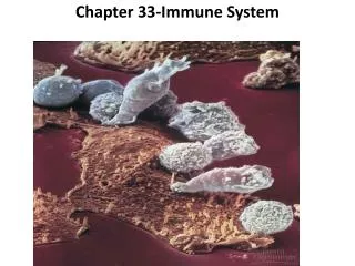
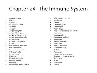
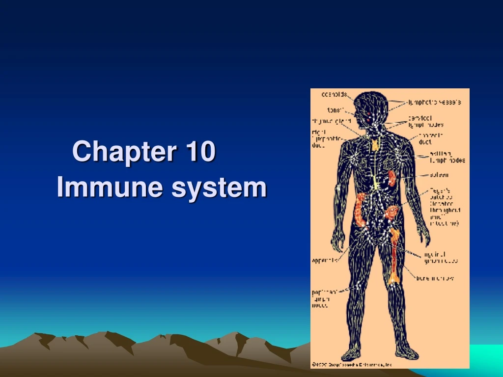
Chapter 10 Immune system. 1. Components. 1) Cells. ① Lymphocyte : a. T-lymphocytes : cytotoxic T cell : Tc C- kill the tumor cell, virus infective cell and foreign cell helper T cell : Th C- promotes activity of BLC and Tc C

E N D
1. Components 1) Cells
① Lymphocyte: a. T-lymphocytes: • cytotoxic T cell: Tc C- kill the tumor cell, virus infective cell and foreign cell • helper T cell: Th C- promotes activity of BLC and Tc C • suppressor T cell: Ts C –regulate the function of BLC and TC b. B-lymphocytes: become into plasma cell c. NK cell: counteract virus infective cell and tumor cell
②Plasma cell ③antigen presenting cell: a. dendritic cell: • Blood DC • Langerhans cell • interstitial cell • veiled cell • interdigitating cell b.macrophage: Mononuclear phagocytic system
④other cells: • granulated cell • mast cell • blood platelet • blood-borne stem cell *Function: i. immunologic defence function ii. immune surveillance function iii. immune homeostasis
2) Lymphoid tissue ---reticular T: • reticular cell: stellate-shaped with processes to form network • reticular fiber ---lymphocytes, macrophage, plasma cell and mast cell
a. Diffuse LT: • no clear boundary • mainly consists of TLC • postcapillary venules: -high endothelial venules -opening for LC enter LT from blood
b. Lymphoid nodule: • spherical or ovoid • have clear boundaries • mainly composed of BLC • germinal center: stained pale * primary LN → secondary LN
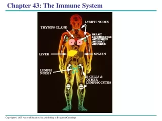
3) Lymphoid organs • Central lymphoid organs: thymus, bone marrow • developed earlier • blood-borne stem cell comes from yolk sac • microenvironment → proliferation promoting • send LC to PLD and LT two weeks before borne
b. Peripheral lymphoid organs: lymph node, spleen and palatine tonsil • developed later • LC come from CLO • cell proliferation need antigen stimulating - antigen dependent • place for immune reactions
2.thymus • Structure of thymus ---capsule: CT→interlobular septum ---cortex: dark-staining ---medulla: lighter-staining ---consists of thymic epithelial cell, thymic stromal cell and thymocytes
1)cortex: ---thymic epithelial cell (epithelial reticular cell): • subcapsular epithelial cell: /flattened /secret β 2-microglobulin, thymosin and thymopoietin • interdigitation dendritic cell: /more processes /MHC antigen
---thymocyte: different stages of LC large LC (prethymocyte): superfacial cortex common thymocyte: deep cortex- 85-90%
2) medulla: ---epithelial reticular cell • medullary epithelial cell: secret thymosin • thymic corpuscle epithelial cell ---thymocyte, macrophage
thymic corpuscle: /spherical or ovoid, 30-50um in D /concentrically-arranged epithelial reticular cells /peripheral cell: inmature /near centra: mature /center cell: keratinased-eosinophilic, hyalinised, with invading of macrophage, eosinophil and LC /function: unknown
3) Blood-thymus barrier: ---Components • contineous endothelial cell • complete basement membrane of endothelium • peri-vessel space containing macrophage • basal lamina of epithelial reticular cell • processes of epithelial reticular cell
---Function provide a stable environment for lymphocytes development
4) Thymus function: a. Place for mature and differentiation of TLC b. Immune regulation
3. Lymph node ---widely distributed ---in groups ---ovoid or kidney-shaped with hilum ---afferent and efferent lymphatic vessel
1) Structure ---capsule: CT, trabeculae or septa ---cortex: outer densely-stained part ---medulla: inner paler-stained part
①Cortex: a. superfacial cortex: ---lymphoid N: • BLC, Macrophage, Th, FDC • primary LN → secondary LN *germinal center: central pale area /dark zone: large, immature BLC, Th /light zone: medium-sized BLC, Th, macrophage, FDC /cap: small BLC ---diffuse LT: thin layer
FDC: (follicular dendritic cell) • light zone • no expression of MHC-II molecules (major histocompatibility complexes) • have Fc receptor and C3 receptor: collect the antigen-antibody complexes and transfer them to BLC and Th C • function: i. active the BLC ii. regulate the synthesis of antibody
b.Paracortex zone-deep cortexunit ---diffuse LT: • TLC, Marcophage, Th cell • interdigitating cell: DC cell -more processes -N: irregular -less organelle -express MHC-II
postcapillary venules: -thick endothelial cell -LC within the wall -opening for LC enter LN
c. Cortical sinus ---subcapsular sinus: afferent LV enter ---peritrabecular sinus structure: • endothelium • RF and RC • cavity: -endothelial cell: stellate for support -macrophage: filter lymph -veiled cell: Langerhans cell ( phagocytose antigen) → efferent LV → LN → paracortex zone
②Medulla ---medullary cord: LT cord: /BLC, plasma cell, macrophage, mast cell /postcapillary venules: channel ---medullary sinus: /similar to cortical sinus and connect with cortical sinus /more macrophage
③Passage of Lymph in LN afferent LV →subcapsular sinuses→peritrabecular sinuses→(narrow channel) →medullary sinus→efferent LV
3) Functions: a.Filter the lymph b.Place to give rise to the immune response c. Involve in the recirculation of LC *Recirculation of LC: • LC (blood) →postcapillary venules → LN → medulla → efferent LV → Blood →LN artery → postcapillary venules →LN • Time: 24-48h
4. Spleen 1) Structure ---capsule: thick DCT with SM and mesothelium, also form trabeculae ---white pulp ---marginal zone ---red pulp
① White pulp: • 1-2mm gray-white spots • periarterial lymphatic sheath: -central artery -diffuse LT : TLC, macrophage, interdigitating cell • splenic corpuscle: -BLC, macrophage, FDC -lymphoid nodules
② Marginal zone: • 100um width • TLC, BLC, macrophage, less erythrocyte • marginal sinus: central artery’s branch- channel for antigen and LC enter LT • place: capture recognize and induce immune reaction
③ Red pulp: a. splenic cord: • LT cord • BLC, DC. M, TC , erythrocytes • place: filter blood b. splenic sinus: • Blood sinus; 12-14um • endothelial cell: rod-liked, gap • RT • basal lamina: incomplete • M-more
2) Blood supply of spleen splenic A→trabecular A→central A branches → marginal sinuses penicillar Arterioles (including: pulp arteriole→ sheathed capillary→ arterial capillary) → splenic sinus→ pulp venule→ trabecular vein→ splenic vein
3) Function: a. filter the blood b. immune reaction c. production of blood in fetus d. blood storage: 40 ml
Tonsil(Study by yourself) ---palatine tonsil ---pharyngeal tonsil ---lingual tonsil
palatine: ---structure: • Stratified squamous epi: invaginated to form many crypts • Epithelium of crypt contain LC, PC, Ma and Langerhans cell • Space and channel between epi.cell: opening to crypt epi. surface, LC filling the channel- lymphoepithelial tissue • Lamina propria: Diffuse LT and LN THE END 谢 谢 !