Download
1 / 36
540 likes | 2.41k Views
Superficial and Cutaneous Mycoses. Eva L.Dizon,MD. Limited to the outermost layer of the skin 4 Infections 1.Pityriasis versicolor 2.Tinea nigra 3. Black piedra 4. White piedra. Superficial Mycoses. Superficial. Do not elicit immune response No discomfort Cosmetic problems

E N D
Superficial and Cutaneous Mycoses Eva L.Dizon,MD
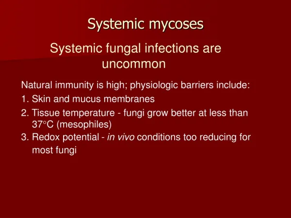
Limited to the outermost layer of the skin 4 Infections 1.Pityriasis versicolor 2.Tinea nigra 3. Black piedra 4. White piedra Superficial Mycoses
Superficial • Do not elicit immune response • No discomfort • Cosmetic problems • Limited to stratum corneum
Pityriasis Versicolor • Malassezia furfur (Pityrosporum orbiculare) • Lipophilic yeast like organism • Rich in sebaceous glands • Media is supplemented with fatty acids • Exist in budding yeast,occasionally hyphal
Pityriasis • Lesions are found in torso, arms and abdomen • Scale very easily chalky appearance • Rarely, papular or grow like folliculitis
Clinical Diagnosis: KOH- Spaghetti and meatballs Treatment: Azoles Pityriasis
Tinea Nigra • Exophiala werneckii • Produce melanin black or brown color • Grows as yeast Older hyphae with mycelia and conidia
Lesion- gray to black macular palms Diagnosis- Skin scrapings with alkali stain Cultures- Sabourauds’s media pigmented yeast and hyphae Tinea nigra
Black Piedra • Piedraia hortae- exist in teleomorphic state • Cultures – asexual state - older cultures teleomorphic (asci ,which contain spindle shaped ascospores)
Black piedra • Clinical feature: presence of hard nodules found along the infected hair shaft • Nodules contain asci
White Piedra • Trichosporon beigelii • Grows in media without cyclohexamide • Cultures are pasty and white developed deep radiating furrows and become yellow and creamy
White Piedra • Microscopic examination septate hypae that develops into arthroconidia • Hair- soft ,pasty,cream colored growth
Treatment • Skin removal of the organism by: 1.Selenium sulfide 2.Thiosulfate 3.Salicylic acid 4.Hyposulfite inhibition of ergosterol by: 1.miconazole
Cutaneous mycoses • Skin • Hair • Nails • Evoke cellular immune response • Dermatophytes • Clinical manifestationsringworm or tinea
Cutaneous mycoses • Etiology Microsporum Trichophyton Epidermophyton
Cutaneous mycoses • Classifications: Anatomic location Tinea pedis Tinea capitis Tinea corporis Tinea cruris Ecologic location Geophilic Zoophilic Anthrophilic
Cutaneous mycoses • Keratophilic – use keratin as subject to live ( parasites) • Keratinases- invade only keratinized layers
2 basic types of dermatophytic infection: 1. The acute or inflammatory type of infection, which is associated with CMI to the fungus, generally heals spontaneously or responds nicely to treatment. 2. The chronic or non-inflammatory types of infection, which is associated with a failure to express CMI to the fungus at the site of infection, is relapsing and responds poorly to treatment.
Cutaneous mycoses • THE IDENTIFICATION REACTION(ID) • Patients infected with a dermatophyte may show a lesion, often on the hands, from which no fungi can be recovered or demonstrated. • It is believed that these lesions, which often occur on the dominant hand (i.e. right-handed or left-handed), are secondary to immunological sensitization to a primary (and often unnoticed) infection located somewhere else (e.g. feet). • These secondary lesions will not respond to topical treatment but will resolve if the primary infection is successfully treated.
Cutaneous mycoses • Laboratory diagnosis: scrapings from clinical specimens • Hair – endothrix (spores inside the hair shaft) -ectothrix - exception: T.schoenleinii Disease-favus-waxy mass of hyphal elements (scutulum) microscopic –degenerated hyphal elements
Cutaneous mycoses • Cultures • Selective media – containing cycloheximide and chlorampenicolincubate at 25 C. • Identification based on the conidia
General characteristics of Macroconidia and Microconidia of Dermatophytes
Diagnosis • Diagnosis is based upon: 1. Anatomical site infected 2. Type of lesion 3. Examination with a Woods lamp (366 A°) 4. Examination of KOH-treated skin scales from the infected area 5. Culture of the organism (not too important)
Differential diagnosis • In a differential diagnosis you must consider: 1. Leprosy 2. Secondary syphilis 3. Pityriasis rosea 4. Psoriasis 5. Nummular eczema 6. Lichen planus 7. Alopecia areata 8. Trichotillomania 9. Dyshidrosis 10. Contact dermatitis.
Treatment • Skin – azoles,inhibits cytochrome 450 dependent enzyme systems at the demethylation step from lanosterol to ergosterol • Hair- Griseofulvin, oral , affects microtubular system