Download
1 / 33
400 likes | 904 Views
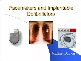
16. Pacemakers and Implantable Cardioverter-Defibrillators. Fast & Easy ECGs, 2nd E – A Self-Paced Learning Program. Artificial Pacemakers. Medical devices used to generate and deliver electrical impulses to the myocardium to stimulate a normal heartbeat

E N D
16 Pacemakers and Implantable Cardioverter-Defibrillators Fast & Easy ECGs, 2nd E – A Self-Paced Learning Program
Artificial Pacemakers • Medical devices used to generate and deliver electrical impulses to the myocardium to stimulate a normal heartbeat • Some are external to the body and provide temporary treatment, others are permanently implanted in the chest
Artificial Pacemakers • One type of temporary pacemaker is the transcutaneous pacemaker • It delivers electrical impulses through lead wires to electrode pads that are applied to the surface of the patient’s chest • Permanent pacemakers are implanted in a surgically created pocket beneath the skin in the patient’s chest wall just below the clavicle
Permanent Pacemakers • Consist of: • A generator • One or more lead wires • A power source (often a lithium battery) • Logic circuits that detect cardiac electrical activity and determine the appropriate response I
Permanent Pacemakers • May be used to: • Symptomatic bradycardia • Sick sinus syndrome • Atrial fibrillation with bradycardia • 3rd-degree (complete) AV heart block • Symptomatic 2nd-degree AV heart block, particularly type II • The sudden development of various combinations of AV heart block and bundle branch block in patients experiencing acute MI • Recurrent tachycardias that can be overdriven and thereby terminated by pacemaker activity • Synchronization of the heart beat in heart failure (cardiac resynchronization therapy)
Pacemaker Electrodes • Are either positioned in the atrium or ventricle alone (single-chamber pacemakers) or, more often, in both chambers (dual-chamber pacemakers or AV sequential pacemakers)
Permanent Pacemakers • Are programmable • Receive and transmit data/programming instructions through the skin using electromagnetic waves • Adjustments can be made to: • Output • Sensitivity • Refractory period • Rate adaption
Pacing Modes • Single-chamber • One pacing lead is inserted into either the right atrium or right ventricle but not both • Dual-chamber • Electrodes are placed into two chambers of the heart • One lead paces the atrium while the other paces the ventricle • By assisting the heart in coordinating the function between the atria and ventricles, this type of pacemaker acts similarly to how the heart naturally paces itself • Also referred to as an AV sequential pacemaker • Most can be programmed to a single chamber mode, which can be useful if the atrial lead wire fails
Pacing Modes • Fixed-rate • Paces the heart at a single, preset rate • Rate-responsive • Has sensors that identify increases or decreases in the patient’s physical activity and automatically adjusts base pacing rate to meet the body’s metabolic needs • Can boost the heart rate in response to motion or increased respirations for those patients whose body cannot appropriately increase the heart rate during activity
Pacing Modes • Demand • Most common type used • Fires only when the patient’s intrinsic heart rate falls below a given threshold level • i.e., if the pacemaker is set at 60 beats per minute it remains inactive until there is a pause between beats that translates into a rate below 60, then the pacemaker fires
Coding System • Pacemaker mode and function described by a five letter coding system • in practice, only three to four are commonly used
Coding System • First letter represents the heart chamber being paced. This letter may be • O = none • A = atrium • V = ventricle • D = dual (ventricle and atrium) • Second letter represents the chamber of the heart being sensed by the pacemaker. This letter may be • O = none • A = atrium • V = ventricle • D = dual (ventricle and atrium)
Coding System • Third letter indicates how the pacemaker generator responds to sensing. This letter may be • O = none • T = triggers pacing • I = inhibits pacing • D = dual (triggers and inhibits pacing) • Fourth letter has to do with adjustment of the pacing rate in response to exercise • If pacemaker is rate responsive, it is denoted with the letter “R” • If there is none, it is denoted as “O”
Coding System • The fifth letter indicates multisite pacing. This letter may be • O = none • A = atrium • V = ventricle • D = dual (ventricle and atrium)
Coding System Examples • VOO • In this mode, the ventricle is paced and there is no sensing function • AAI • Pacemaker paces and senses in the atrium • When it senses atrial activity, pacing is inhibited • VVI • Ventricle is paced and sensed • If spontaneous cardiac output is detected, then the device is inhibited
Coding System Examples • VDD • Here the pacemaker paces the ventricle and senses both the atrium and ventricle • On sensing intrinsic atrial activity, the pacemaker triggers ventricular pacing; on sensing ventricular activity, the pacemaker inhibits pacing • It is also known as a P-synchronous pacer
Coding System Examples • DVI • Pacemaker can pace in the atrium, the ventricle, or both • Sensing takes place only in the ventricle • When the pacemaker senses intrinsic ventricular activity, it inhibits pacing • DDD • Pacemaker paces and senses in the atrium, the ventricle, or both • On sensing activity in either chamber, the pacemaker inhibits pacing in that chamber • Or, on sensing atrial activity, the pacemaker may trigger ventricular pacing
Cardiac Resynchronization Therapy (CRT) • Used to resynchronize a heart that does not beat in synchrony, a common problem in patients with heart failure • Employs three leads: • one is placed in right atrium • one is located in right ventricle • last one is inserted through the coronary sinus to pace the free wall of the left ventricle • These three wires are connected to a CRT generator and programmed so that the two ventricular wires are activated simultaneously I
Unipolar and Bipolar Systems • Unipolar • positive electrode is positioned in the heart tissue and the negative electrode is connected to the pulse generator • produces tall pacing spikes on the ECG • In a bipolar system, • electrodes are only millimeters apart in the cardiac tissue • produces short pacemaker spikes I
ECG Features of a Pacemaker • Depending on how many chambers are paced, the firing of a pacemaker produces one or two narrow pacemaker spikes on the ECG
ECG Features of a Pacemaker • A paced ECG complex shows two features: (a) a narrow “pacing spike,” which reflects the impulse depolarizing the paced chamber and (b) a P wave or QRS complex that immediately follows the pacing spike I
Pacemaker Failure • Pacemakers may not work properly for a number of reasons, including a failure to capture, a failure to pace, a failure to sense, oversensing, and pacemaker-mediated tachycardia
Pacemaker Failure • Failure to capture is seen as the presence of pacemaker spikes that are not followed by a P wave or broad QRS complex
Pacemaker Failure • Failure of the pacemaker to sense is seen as the presence of ECG pacemaker spikes that fall where they shouldn’t
Pacemaker Failure • Oversensing is seen as an absence of pacemaker spikes in the presence of a heart rate that is slower than the rate set for the pacemaker
Pacemaker Failure • Pacemaker-mediated tachycardia is seen as a fast heart rate with a pacemaker spike preceding each QRS complex on EGG.
Implantable Cardioverter-Defibrillator (ICD) • Is implanted in patients who are at risk of sudden cardiac death due to ventricular fibrillation and ventricular tachycardia
Implantable Cardioverter-Defibrillator • Is programmed to detect cardiac dysrhythmias and correct them by delivering paced beats, cardioversion, or defibrillation
Practice Makes Perfect • Analyze this ECG tracing I
Practice Makes Perfect • Analyze this ECG tracing I
Practice Makes Perfect • Analyze this ECG tracing I
Practice Makes Perfect • Analyze this ECG tracing I