Download
1 / 20
200 likes | 324 Views
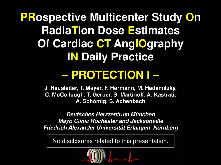
PR ospective Multicenter Study O n Radia T ion Dose E stimates Of Cardiac CT Ang IO graphy I N Daily Practice – PROTECTION I –

E N D
PRospectiveMulticenter StudyOnRadiaTionDoseEstimatesOf CardiacCTAngIOgraphy INDaily Practice – PROTECTION I – J. Hausleiter, T. Meyer, F. Hermann, M. Hadamitzky, C. McCollough, T. Gerber, S. Martinoff, A. Kastrati, A. Schömig, S. AchenbachDeutsches Herzzentrum MünchenMayo Clinic Rochester and JacksonvilleFriedrich Alexander Universität Erlangen–Nürnberg No disclosures related to this presentation.
Background • Cardiac CT angiography (CCTA) has evolved as a useful non-invasiveimaging modality. • With its rapid increase in use, the exposure to ionizing radiation associated with cardiac CT angiography has raised serious concerns.
Rationale to determine in a prospective study: • the radiation dose of cardiac CT angiographies in daily practice • the efficacy of dose saving algorithms • independent predictors associated with radiation dose
Methods I • Study design: prospective, observational multi-center multi-vendor industry-independent • Enrollment period: Feb. – Dec. 2007 • 50 participating study sites1965 CCTAs • Image data, patient and scan information of all consecutive ECG-gated or -triggered CCTAs performed during one month
Methods II • Calculation of estimated radiation dose:European Working Group for Guidelines on Quality Criteria in CT Dose-length-product (DLP) * 0.017 (conversion factor chest) • Image quality:Diagnostic image quality assessed on a per-vessel basis (diagnostic vs. non-diagnostic) • Linear regression analysis:Identification of independent factors influencing dose
50 participating study sites Argentina Vicente Lopez, P. Carrascosa, A. Deviggiano Australia Frankston, G. Szto, A. Watson Austria Innsbruck, G. Feuchtner, G. Friedrich Belgium Aalst, P. Vanhoenacker, I. Decramer Brussels, E. Coche, B. Gerber Antwerp, B. Shivalkar, R. Salgado Brazil Sao Paolo, R. Sasdelli Neto, I. Pinto Rio de Janeiro, A. Oliveira, D. M. Moreira Canada Montreal, C. Chartrand-Lefebvre, J. Prenovault Vancouver, B. Forster, D. Malpas Denmark Aarhus, O. Gøtzsche, E. Morre Pedersen Germany Bad Krotzingen, J. Allgeier, F.-J. Neumann Bad Nauheim, T. Dill, J. Rixe Bad Oeynhausen, C. Langer, D. Horstkotte Erlangen, S. Achenbach, T. Pflederer Essen, O. Bruder, T. Schlosser Frankfurt, A. Schmermund, A. Magedanz Kiel, T. Jahnke, T. Huemme Landshut, E. Sauer, J. Dietl Munich, C. Becker, A. Leber Munich, R. Haberl, G.-E. Böhme Munich, J. Hausleiter, S. Martinoff Rosenheim, M. Block, C. Baierl Saarbrücken, G. Goerge, J. Schmitt Traunstein, W. Moshage, A. Opitz Ulm, M. Hoffmann, O. Klass Great Britain London, C. di Mario, N. Arcuri Middlesex, T. Mittal, T. Patel Greece Athen, I. Mastorakou, T. Syrigou Israel Haifa, O.-R. Brook, S. Abadi Italy Rome, E. Martuscelli, E. Casciani Japan Hiroshima, T. Kitagawa, J. Horiguchi Tokyo, S. Kuribayashi, M. Yamada Tokyo, N. Yokoyama, S. Suzuki Korea Seoul, J.-W. Kang, J.B. Seo Malaysia Sarawak, T. Ong, K.-H. Sim Mexico Mexico City, E. Alexanderson, A. Meave Monterrey, E. de la Pena-Almaguer, R. Zamarripa-Morales Monaco Monaco, F. Civaia, P. Rossi the Netherlands Groningen, M. Greuter, M. Oudkerk Spain Oviedo, C. Paz, J.F. Villameytide Pakistan Karachi, R. Ahmed, S. Kureshi Portugal Vila Nova de Gaia, N. Bettencourt de Sousa, V. G. Ribeiro Singapore Singapore, K.-T. Ho, G. Kaw Spain Malaga, E. Gonzalez Cocina, A. Ruiz Switzerland Zurich, H. Alkadhi, P. Stolzmann Turkey Erzurum, M. Kantarci, F. Fil USA Iowa City, E.J.R. van Beek, J.M. Wilson Fairfax, J.P. Earls, E. Berman Washington, A. Taylor, P.J. Devine
Study site characteristics * Excluded in analysis of dose saving algorithms and linear regression model
Estimated radiation dose Dose (mSv) 50 40 30 20 10 0 15.4 [9.8; 22.0]
Estimated radiation dose Dose (mSv) Range of medians: 5.7 to 36.5 mSv 50 40 30 20 10 0 Study sites
Dose saving algorithms • Automatic exposure controladaption of tube current to pat.‘s anatomy • ECG pulsingmodulation of tube current to pat.‘s ECG • 100 kV tube voltageinstead of conventional ≥ 120 kV tube voltage • Sequential scanning (step and shoot)instead of conventional spiral scan technique
Automatic exposure control– 64-slice systems – Estimated dose 30 (mSv) 20 10 15.4 15.8 0 Frequency of use 100 (%) 50 62.1 37.9 0 without with automatic exposure control
ECG pulsing– 64-slice systems – Estimated dose 30 (mSv) 20% 20 10 20.9 16.7 0 Frequency of use 100 (%) 50 21.3 78.7 0 without with ECG pulsing
100 kV tube voltage– 64-slice systems – Estimated dose Image quality 30 100 (mSv) (%) 20 50% 10 50 8.7 97.3 17.4 97.3 0 0 Frequency of use 100 (%) 50 5.8 94.2 0 ≥ 120 100 kV tube voltage
Spiral vs. sequential scanning– 64-slice systems – Estimated dose Image quality 30 100 (mSv) (%) 20 68% 10 50 17.6 5.6 97.2 98.3 0 0 Frequency of use 100 (%) 50 6.2 93.8 0 spiral sequential scanning
Predictors of radiation dose– in 64-slice systems – Weight ( 10kg) <.0001 Heart rhythm (sinus vs. non-sinus) 0.0002 Heart rate ( 10bpm) 0.58 Indication (coronary vs. non-coronary) 0.98 Scan length ( 1cm) CT system (highest vs. lowest) <.0001 <.0001 100 vs. ≥120kV tube voltage <.0001 Sequential scanning ECG pulsing <.0001 <.0001 Site experience ( 1 year) 0.005 -25 -20 -15 -10 -5 0 5 10 15 Effect in linear regression analysis [mSv]
Predictors of radiation dose- Impact of CT system - GE 64 Toshiba 64 Siemens dual-source 64 Philips 64 Siemens single-source 64 0 2 4 6 8 10 12 14 Effect in linear regression analysis [mSv]
Radiation dose from cardiac CT angiography varies significantly between study sites and CT systems. Although very effective measures to reduce the radiation dose are available (100 kV and sequential scanning),these are rarely used in daily practice. Conclusion I
Worldwide educational efforts (by medical societies & CT vendors) are mandatory to ensure the uniform and consistent use of dose saving algorithms where applicable. Further developments and critical evaluations of additional strategies for radiation dose savings are needed. Conclusion II