Download

1 / 49
990 likes | 2.15k Views
Familial Hypercholesterolemia. KEY POINTS . FH is an autosomal dominant genetic condition that leads to severe elevations in cholesterol levels. Average LDL is 220mg/dl in HeFH and > 500mg/dl in HoFH

E N D
KEY POINTS • FH is an autosomal dominant genetic condition that leads to severe elevations in cholesterol levels. • Average LDL is 220mg/dl in HeFH and > 500mg/dl in HoFH • Lifetime burden of high cholesterol leads to huge increased risk of cardiovascular disease. • 20 fold increased risk of premature cardiovascular disease (CVD) • FH is among the most common inherited conditions: • Prevalence of heterozygous FH (HeFH) is 1:300-1:500. • Homozygous FH is rare at 1:1,000,000 but has terrible consequences • FH affects all race/ethnic groups • FH can be diagnosed based on a combination of lipid levels, family history, physical exam findings and genetic testing. • FH is massively underdiagnosed and undertreated. • There are > 600,000 people in the US with FH but only 10% have been diagnosed. • FH is treatable • With statin-based therapy, risk of CVD can be greatly reduced • Because of the genetic nature of the disease, once one person in a family is diagnosed with FH, it is mandatory to screen the rest of the family members (so called “cascade screening”)
OVERVIEW Familial Hypercholesterolemia
Overview of FH • FH is almost always inherited as an autosomal dominant disorder with a very rare autosomal recessive form1,2 • Low-density lipoprotein cholesterol (LDL-C) generally exceed the 95th percentile during childhood in patients with FH2,3 • LDL-C concentrations are generally 2- to 3 fold higher in people with HeFH and 3- to 6-fold higher than normal in HoFH.4 • Patients with FH have heightened risk of CVD due to life-long exposure to elevated cholesterol 5,6 • If left untreated, individuals with HoFH often develop symptomatic CVD before age 25. Those with untreated HeFH often experience the same by age 55.1 • A small fraction of treated FH patients have cardiovascular disease (CVD) and persistently elevated LDL-C levels >200 mg/dL (5.17 mmol/L) or have no CVD but persistently elevated LDL-C levels >300 mg/dL (7.75 mmol/L) despite maximally tolerated lipid-lowering therapy. These FH patients are referred to as severe FH and are currently eligible for LDL apheresis in the United States. • Marais AD. Clin Biochem Rev. 2004;25(1):49-68. • Mahley RW, et al. In: Kronenberg: Williams Textbook of Endocrinology; 2008. • Graaf et al . Circulation. 2011;123:1167-1173.). • 4. Vella A, et al. Mayo Clin Proc. 2001;76(10):1039-1046. 5. Goldberg AC, et al. J Clin Lipidol. 2011:5:S1-S8. 6. Rader DJ, et al. J Clin Invest. 2003;111(12):1795-1803. 7. Thompsen J, Thompson PD. Atherosclerosis. 2006;189:31-38.
Table adapted from Vella A, et al. Mayo Clin Proc. 2001;76:1039-1046. • Thompsen J, Thompson PD. Atherosclerosis. 2006;189:31-38. • Raal FJ, et al. Atherosclerosis. 2000;150(2):421-428. • 3. Ito MK, et al. J Clin Lipidol. 5(3):S38-S45.
FH is due to impaired LDL clearance and increased secretion of ApoBparticles Atherosclerosis and tissue damage Increased plasma LDL Impaired LDLR-mediated clearance Increased LDL uptake bynon-LDLR-mediated pathways Decreased degradation of ApoB and increased production and secretion of ApoB-containing lipoproteins into the circulation Figure adapted from data and concepts in adjacent references. 1. Barrett PH, Watts GF. Atheroscler Suppl. 2002;2(3):1-4. 2. Sniderman AD, et al. Clin Sci (Lond). 2009;118(5):333-339. 3. Fisher WR, et al. Arterioscler Thromb. 1994;14(4):501-510. 4. Cummings MH, et al. Atherosclerosis. 1995;113(1):79-89. 5. Tremblay AJ, et al. J Lipid Res. 2004;45(5):866-872. 6. Twisk J, et al. J Clin. Invest. 2000;105(4):521-532.
GENETIC BASIS Familial Hypercholesterolemia
Dd Dd FHis almost always autosomal dominant • Most common FH-causing mutations are in the LDL receptor gene (LDLR). Less common defects include mutations in APOBor PCSK9 genes1 • Heterozygotes inherit a single abnormal gene from one parent, Given the dominant mode of inheritance, these individuals manifest the disorder.2 • Heterozygotes have approximately 2- to 3-fold higher serum LDL-cholesterol levels than normal2 • Homozygotes inherit an abnormal gene from both parents. They typically have an LDL- cholesterol level 3- to 6-fold higher than normal2 Autosomal Dominant LDLR, ApoB, and PCSK9 mutations Affected orpredisposedmother with autosomaldominant faulty gene Affected orpredisposedfather with autosomaldominant faulty genes Affected orpredisposedfather with autosomaldominant faulty genes D d d D Eggs Sperm dd Dd Dd DD Unaffected Affected Severely affected 1 out of 4 chances 25% 2 out of 4 chances 50% 1 out of 4 chances 25% A small spectrum of affected heterozygotes may have unusually severe phenotypes; unusually severe phenotypes may also be seen in compound heterozygotes that have mutations in 2 different genes (1 from each parent or both from the same parent)3,4 • Marais AD. Clin Biochem Rev. 2004;25(1):49-68. • Vella A, et al. Mayo Clin Proc. 2001;76(10):1039-1046. • Pisciotta L, et al. Atherosclerosis.2006;186(2):433-440. • Tai ES, et al. Clin Chem. 2001;47(3):438-443. Image adapted from www.genetics.edu.au/pdf/factsheets/fs09.pdf.
FHis very rarely autosomal recessive Autosomal Recessive ARH/LDLRAP1 mutations • Mutations in the LDLRAP1 gene result in a very rare autosomal recessive form of homozygous FH called autosomal recessive hypercholesterolemia (ARH)1 • The clinical phenotype of homozygous ARH is similar to that of classic homozygous familial hypercholesterolemia but it is somewhat milder in terms of serum total cholesterol and LDL-C levels1,2 • Because it is a recessive disorder, most ARH patients are homozygous for the same allele inherited from consanguineous or related parents1 • Because ARH is an autosomal recessive disorder, the heterozygotes are unaffected1 Unaffected geneticcarriermother Unaffectedgeneticcarrierfather Rr Rr Sperm r R Eggs r R rr Rr Rr RR Non-carrier (unaffected) Geneticcarriers (unaffected) Severely affected 1 out of 4 chances 25% 2 out of 4 chances 50% 1 out of 4chances 25% Image adapted from www.genetics.edu.au/pdf/factsheets/fs09.pdf. • Soutar AK, et al. Arterioscler Thromb Vasc Biol. 2003;23(11):1963-1970. • Pisciotta L, et al. Atherosclerosis. 2006;188(2):398-405.
FH can be caused by mutations in 4 known genes FH is typically caused by mutations in LDLR, ApoB, PCSK9, LDLRAP1 or other as yet other unidentified genes1 Apo B acts as ligand, binding LDL particle to receptor LDL Particle Circulation Liver cell LDL Receptor on hepatocyte, binds to Apo B on LDL particle, inducing endocytosis of LDL LDLRAP1 (ARH) mediates internalization via clathrin coated pits PCSK9 Enzyme degrades LDL receptors Image reproduced from http://www.dls.ym.edu.tw/ol_biology2/ultranet/Endocytosis.html. 1. De Castro-Oros I, et al. Appl Clin Genet. 2010;3:53-64.
FH-related mutations Phenotype of FH Table adapted from Rader DJ, et al. J Clin Invest. 2003;111:1795-1803 and De Castro-Oros I, et al. Appl Clin Genet. 2010;3:53-64. • De Castro-Oros I, et al. Appl Clin Genet. 2010;3:53-64. • Noguchi T, et al. Atherosclerosis. 2010;210(1):166-172. • Soutar AK, et al. Arterioscler Thromb Vasc Biol. 2003;23(11):1963-1970. • 4. Rader DJ, et al. J Clin Invest. 2003;111(12):1795-1803. • Zulewski H, et al. J Lipid Res. 1998;39(2):380-387. • Ouguerram K, et al. Arterioscler Thromb Vasc Biol. 2004;24(8):1448-1453.
PATHOPHYSIOLOGY Familial Hypercholesterolemia
The atherosclerotic burden of FH reflects the lifelong accumulation of LDL and all ApoB-containing lipoproteins ApoB 100 ApoB- containing lipoproteins: Non-HDL-C:plasma cholesterol concentration of ApoB-containing particles Apo(a) Lp(a) VLDL IDL LDL Cholesterol concentration measures: LDL-C Non-HDL-C Adapted from Robinson JG*J Am Coll Cardiol. 2010;55(1):42-44.
LDL accumulates to cause CHD early in life in FH Age Patients Meet CHD Threshold HoFH Normal HeFH 10 Female Threshold for CHD Male Hypertension Diabetes Smoking Cumulative LDL-C (g/dL-years) 5 0 1 20 40 60 80 Age (years) • Threshold for CHD reached by: • Age 15 y in HoFH patients • Age 40 y in HeFH patients • Age >60 y in healthy individuals Adapted from Horton JD, et al. J Lipid Res. 2009;50(Suppl):S172-S177.
Arterial Intima-media Thickness in children with FH • Significant differences in intima-media thickness (IMT) were noted by age 12 in children with FH versus unaffected siblings (P = 0.002) 0.08 0.06 Child with FH 0.04 FH – control IMT (mm) 0.02 Significant difference in IMT Unaffected sibling 0 –0.02 –0.04 8 10 12 14 16 18 Age (y) Adapted from Wiegman A, et al. Lancet. 2004;363(9406):369-370.
Carotid IMT in FH versus non-FH patients 1.6 1.4 FH patients: ~45 y Healthy non-FH: ~80 y 1.2 FH individuals 1.0 cIMT (mm) Linear (pooled FH individuals) 0.8 Non-FH individuals 0.6 Linear (pooled non-FHindividuals) 0.4 10 20 30 40 50 60 70 80 Age (y) Adapted from De Groot E, et al. Circulation. 2004;109[ suppl III]:III33–III38.
Xanthelasma Tendon xanthomas Arcus cornealis LDL-C concentrations and physical signs in untreated FH patients Cumulative Prevalence of Physical Signs in Adult FH Patients3 LDL-C Distribution in Untreated Patients With Definite FH3 Median = 6.95 mmol/L (268.76 mg/dL) Mean = 7.22 mmol(279.2 mg/dL) 100 75 Cumulative prevalence, % 50 Relative frequency 25 0 15 25 35 45 55 65 75 85 3.0 3.5 4.0 4.5 5.0 5.5 6.0 6.5 7.0 7.5 8.0 8.5 9.0 9.5 10.0 10.5 11.0 11.5 12.0 12.5 13.0 13.5 14.0 14.5 15.0 15.5 LDL-C (mmol/L) in untreated patients Age (y) 1. Marais AD. Clin Biochem Rev. 2004;25(1):49-68. 2. Raal FJ, et al. Atherosclerosis. 2000;150(2):421-428. 3. Blom DJ, et al. S Afr Fam Pract. 2011;53(1):11-18.
EPIDEMIOLOGY Familial Hypercholesterolemia
The prevalence of HeFH is ~1:5001-3 The prevalence of HoFH is ~1:1,000,0002,3 FH is a common inherited disorder 2.0 per 1000 1.0 0.8 0.5 0.5 0.5 0.5 0.4 0.4 0.3 FH Sickle cell disease Multiple exostoses Huntington's disease Fragile X syndrome Neuro-fibromatosis Cystic fibrosis Duchenne muscular dystrophy Adult PCKD Dominant otosclerosis PCKD, polycystic kidney disease. Figure adapted from Genetic Alliance UK. Available at http://www.geneticalliance.org.uk/education3.htm. • Citkowitz E. Familial Hypercholesterolemia. http://emedicine.medscape.com/article/121298-overview#a0199. • Vella A, et al. Mayo Clin Proc. 2001;76(10):1039-1046. • Austin MA, et al. Am J Epidemiol. 2004;160(5):407-420.
In some populations FH is even more common Founder Populations 1:67 1:72 to 1:100 1:165 1:85 1:270 1:500 General population Adapted from Austin MA, et al. Am J Epidemiol. 2004;160(5):407-420.
CLINICAL PRESENTATION Familial Hypercholesterolemia
Heterozygous FH • 20-fold increased risk of CVD • If untreated: • Men have 50% risk of CVD by age 50 • Women have 30% risk of CVD by age 60 • Many HeFH patients present with established CVD (angina, MI)
A B C D E F Physical exam findings in FH A. Xanthelasma B. Corneal arcusa C. Achilles tendon xanthomas D. Tendon xanthomasb,1-3 E. Tuberous xanthomasc F. Planar xanthomasc • A Common in older individuals (even non-FH); definitive of FH in younger individuals. • B 30%-50% of the HeFH population have tendon xanthomas. • C Seen mostly in HoFH, and not as often in HeFH • Ferrières J, et al. Circulation. 1995;92(3):290-295. • Bertolini S, et al. Arterioscler Thromb Vasc Biol. 2000;20(9):E41-E52. • Descamps OS, et al. Atherosclerosis. 2001;157(2):514-518. Figure adapted from Mahley RW, et al. In: Kronenberg HM. Williams Textbook of Endocrinology. 11th ed. Philadelphia: Saunders; 2008.
Clinical presentation of HoFH • Cutaneous xanthomas at birth or by early childhood1 • Planar xanthomas(on hands, elbows, buttocks, or knees), which are diagnostic for the homozygous state1 • Tuberous xanthomas(on hands, elbows, or knees)1 • Tendon xanthomas (especially on extensor tendons of hands or Achilles tendon)1 • Valve affection murmur of aortic stenosis may be heard1,2 A Six-year-old girl with HoFH. Bumps on skin are deposits of cholesterol derived from LDL. B C • Image A reproduced from Brown/Goldstein Lab. Department of Molecular Genetics at UT Southwestern. http://www4.utsouthwestern.edu/moleculargenetics/pages/gold/past.html. • Image B reproduced from Li SG, et al. N Engl J Med. 2009;360(18):1885. • Image C reproduced from Thappa DM, Karthikeyan K. Indian Pediatr. 2003;40(6):574-575. • Citkowitz E. Familial Hypercholesterolemia. http://emedicine.medscape.com/article/121298-overview#a0199. • Allen JM, et al. Br Heart J. 1980;44(4):361-368.
HoFH clinical course • Severe vascular disease at an early age. • Numerous case reports of CABG or death before age 10. • Severe aortic stenosis common • Untreated, usually results in death before age 30. • Almost all HoFH patients require LDL apheresis but even with maximal medical therapy there is disease progression.
DIAGNOSIS AND SCREENING Familial Hypercholesterolemia
The diagnosis of FH is largely based on Extreme hypercholesterolemia early in life (LDL-C ≥190 mg/dL [adults] or ≥160 mg/dL [children or adolescents])1 Clinical evidence of premature CVD and/or family history of hyperlipidemia2 According to National Lipid Association (NLA) criteria, FH should be suspected inthe following cases1 Children, adolescents, young adults (<20 y) with LDL-C ≥160 mg/dL or non-high-density lipoprotein cholesterol (HDL-C) ≥190 mg/dL Adults (≥20 y) with LDL-C ≥190 mg/dL or non-HDL-C ≥220 mg/dL Formal diagnosis through MEDPED, Simon-Broome or Dutch criteria LDL-C categories often used for FH diagnosis • Goldberg AC, et al. J Clin Lipidol. 2011:5(3 Suppl):S1-S8. • Rader DJ, et al. J Clin Invest. 2003;111(12):1795-1803. • Hopkins PN, et al. J Clin Lipidol. 2011;5(Suppl 3):S9-S17. aThis category is relevant for diagnosis of FH patients who are first-degree relatives of a knownFH case. At the LDL-C level shown, ~ 80% of first-degree relatives can be expected to have FH.
High cholesterol screening recommendations from the NLA • Universal screening for serum cholesterol levels is recommended • Cholesterol screening should be considered starting at age 2 for children with a family history of premature CVD or high cholesterol. • All children should be screened between the ages of 9 – 11 regardless of family history. • For all children with an LDL-C ≥160 mg/dL after adequate lifestyle interventions lipid altering medications are recommended. • “Cascade screening” of all first-degree relatives of diagnosed FH patients should be performed; newly identified FH cases may reveal additional relatives who should be screened Goldberg AC, et al. J Clin Lipidol. 2011:5(3 Suppl):S1-S8.
Genetic testingfor FH • In the US, the diagnosis of FH is usually made on clinical grounds. • However, genetic testing is the gold-standard for the diagnosis of FH and is widely used in many countries. • Genetic testing can be used in cases where the diagnosis is unclear. • Testing generally involves sequencing 3 genes (LDLR, APOB and PCSK9). • The yield on genetic testing is ~85% in “definite” FH cases and ~50% in “possible” FH. • As genetic testing becomes cheaper it is probable that this method will be used more widely. • May be particularly useful for cascade screening.
The CDC has classified genetic testing for FH as a “Tier 1” application (Best evidence to support use)
CURRENT TREATMENT Familial Hypercholesterolemia
Management • Diet/lifestyle changes important • Reduce saturated fat • High soluble fiber: 10-20g/day • Dietician referral • Statins are the mainstay of therapy • Many will require 2 or more drugs • Goal to reduce LDL ≥ 50% (or LDL < 100 ideal) • Treat other risk factors (HTN, smoking) • DO NOT USE STANDARD RISK ASSESSMENT TOOLS TO ESTIMATE RISK • Framingham score is not valid in FH patients
Goals of therapy • FH patients are considered “high risk” based on NCEP, AHA/ACC guidelines • Rule of thumb to try to cut LDL by 50% • Ultimate goal LDL < 100 • Many would try to get LDL < 70 • Unlike “garden variety” HLD, non-statin agents are standard of care. • With proper therapy can reduce risk of CHD by 80%. NCEP ATP Guidelines, Circ 2002
Current therapeutic options for FH: statins Statins competitively inhibit HMG CoA reductase, reducing endogenous cholesterol synthesis LDL Lipid core-cholesteryl esters ApoB Statin LDL receptor Reduced cholesterol synthesis Fatty Acids Acetyl-CoA HMG CoA = Cholesterol ACAT2 Cholesterol Esters MTP ApoB-100 VLDL LDL ApoB- 100 VLDL LDL Triglycerides ApoB- 100 ApoB- 100 Increased receptor expression Increased ApoB-LDL uptake Figure adapted from Stancu C, Sima A. J Cell Mol Med. 2001;5(4):378-387.
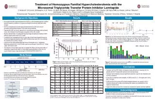
Delayed CV events and prolonged survival in HoFH patients have altered the disease spectrum of HoFH from a fatal disease in childhood to that seen in untreated HeFH Effect of statins and lipid-lowering therapy in FH • Statins have demonstrated significant MI-free survival benefits in HeFH patients Figure 1 Figure 2 A. 100 Benefit From Lipid Therapy (Endpoint: Death) 1.0 Yes 0.8 No 80 0.6 Survival probability 60 0.4 Cumulative event-free survival (%) 0.2 40 Statin treatment HeFH 0 No statin treatment HeFH 0 10 20 30 40 50 60 70 20 Age (y) No statin treatment –general population B. 0 1.0 Benefit From Lipid Therapy (Endpoint: MACE) 0 2.5 5.0 7.5 10.0 12.5 Yes 0.8 Follow-up (y) No 0.6 Survival probability 0.4 0.2 0 0 10 20 30 40 50 60 Age (y) Figure 1 adapted from Versmissen J, et al. BMJ. 2008;337:a2423. Figure 2 adapted from Raal FJ, et al. Circulation. 2011;124(20):2202-2207.
In FH patients, statins are highly effective but the LDL-cholesterol responsiveness to statins is influenced by the nature of the LDL receptor gene mutation and some LDLR mutations tend to render statins less effective4,5 Because most current therapies directly increase LDL-C clearance via the LDLR or decrease LDL-C via other clearance mechanisms that secondarily up-regulates the LDLR, their effectiveness in FH patients can be limited Current therapies with the exception of niacin, do not significantly impact LDL/ApoB production Mechanism of action of current therapies Table adapted from Radar DJ, et al. J Clin Invest. 2003;111(12):1796-1803. Chaves et al. lin Endocrinol Metab 86: 4926–4932,2001 Gordon BR, et al. Am J of Card. 1998;81(4):407-411. Ito MK, et al. J Clin Lipidol. 2011;5(3 Suppl):S38-S45. • Kastelein JJ, et al. N Engl J Med. 2008;358(14);1431-1443. • Raal FJ, et al. Atherosclerosis. 2000;150(2):421-428. • Konrad RJ, et al. Lipids Health Dis. 2011;10:38. • Vohl et al, Atherosclerosis 160 (2002) 361–368
For the worst affected FH patients LDL apheresis is added Cholesterol Rebound After Receiving LDL Apheresis • LDL-C reductions withapheresis: • Acute: Up to 76%1 • Time averaged: 20-40%2 LDL-C (mg/dL) 1 2 3 4 5 6 7 8 9 10 Treatments Reproduced from Thompson GR. Curr Opin Lipidol. 2010. 1. Gordon BR, et al. Am J of Card. 1998;81(4):407-411. 2. Ito MK, et al. J Clin Lipidol. 2011;5(3 Suppl):S38-S45.
Treatment algorithm for the worst affected FH patients • All HeFH patients require lipid-lowering drugs to reach target LDL-C levels1,2 • Adult Treatment Panel (ATP) III guidelines indicate that most FH patients will require combination therapy1,2 • LDL apheresis and/or liver transplant is only required in rare cases1,2 Lifestyle changes Proper diet, reduction in body weight if overweight, smoking cessation, and exercise Statin treatment Combination therapy HoFH and severe FH patients LDL apheresis or liver transplant Schematic adapted from adjacent references 1 - NIH. NCEP, Final Report. Pub No. 02-5215. September 2002.. 2 – Ito et al. Management of Familial Hypercholesterolemias in adult patients: Recommendations from the National Lipid Association Expert Panel on Familial Hypercholesterolemia Journal of Clinical Lipidology (2011) 5, S38–S45
ADDITIONAL RISK FACTORS TO CONSIDER Familial Hypercholesterolemia
Additional CVD risk factors to consider in HeFH • Independent CVD risk factors3,4 • In FH, independent CVD risk factors couldpotentially interact with lifelong high LDL-Clevels to compound risk5 • Lipoprotein(a) [Lp(a)] ≥50 mg/dL1,3,4 • Tendon xanthomas1-3 • Men: ≥30 y1 • Women: ≥45 y or postmenopausal1 • Cigarette smoking: active smokers1 • Family history of premature CVD1 • CVD in male first-degree relative <55 y or in female first-degree relative <65 y • HDL-C <40 mg/dL (1.0 mmol/L)1 • Blood pressure >140/90 mmHg1 • Diabetes mellitus1 • Holmes DT, et al. Clin Chem. 2005;51(11):2067-2073. • Mbewu AD, et al. Arterioscler Thromb. 1991;11(4):940-946. • Goldberg AC, et al. J Clin Lipidol. 2011:5(2 Suppl):S1-S8. • Neil HA, et al. Atherosclerosis. 2003;170(1):73-78. • Civeira F, et al. Arterioscler Thromb Vasc Biol. 2005;25(9):1960-1965.
Risk factors in FH independent of LDL-C • Tendon xanthomas • Tendon xanthomas in FH are associated with CV risk independently of the LDLR gene mutation1 • Approximately 30%-50% of heterozygous FH patients with genetic diagnosis have tendon xanthomas2-4 • Lipoprotein(a) • Elevated serum Lp(a) concentrations may be regarded as a component of the clinical syndrome of FH5 • In homozygous or heterozygous FH, mutations in the LDLR show clear gene-dose effect on Lp(a) plasma levels6 • Lp(a) is an independent risk factor for CVD in heterozygous FH7 • Potential interactions between high plasma concentration of Lp(a) as seen in FH and additional risk factors for CVD (such as elevated life long accumulations of LDL-c as seen in FH) may also potentiate the very high CVD risk of FH patients5 • Civeira F, et al. Arterioscler Thromb Vasc Biol. 2005;25(9):1960-1965. • Ferrières J, et al. Circulation. 1995;92(3):290-295. • Bertolini S, et al. Arterioscler Thromb Vasc Biol. 2000;20(9):E41-E52. • Descamps OS, et al. Atherosclerosis. 2001;157(2):514-518. • Mbewu AD, et al. Arterioscler Thromb. 1991;11(4):940-946. • Kraft HG, et al. Arterioscler Thromb Vasc Biol. 2000;20(2):522-528. • Holmes DT, et al. Clin Chem. 2005;51(11):2067-2073.
Lp(a): An independentandcausalriskfactor • Lp(a) consists of an LDL-like particle and the specific Apo(a), which is covalently bound to the ApoB of the LDL-like particle1,3 • BecauseApo(a) isstructurallyhomologoustoplasminogen, Lp(a)1,3 • Competitivelyinhibitsplasmingeneration – antifibrinolytic1,3 • Can bind toplasminandfibrinogen, promoting atherosclerosis1,3 • Depositsoxidizedphospholipids, increasinginflammationleadingto atherosclerosis1,3 • Promotesplaqueinflammationand instability1,3 • Lp(a) has a causalrelationshiptoincreased CV risk2andisrecognizedtopredictatherosclerosis, MI1 • 2011 NLA Expert Panel citedLp(a) as an independentdriverofvery high risk in FH4 Apo(a) LDL particle ApoB-100 Lipoprotein(a) 1. Kiechl S, Willeit J, J Am Coll Cardiol. 2010;55(19):2168-2170.2. Clarke R, et al. N Engl J Med. 2009;361(26):2518-2528. 3. Kathiresan S. N Engl J Med. 2009;361(26):2573-2574. 4. Goldberg AC, et al. J Clin Lipidol. 2011;5(3 Suppl):S1-S8. Figure adapted from Brown WV, et al. J Clin Lipidol. 2010;4(4):240-247.
In FH, Lp(a) levels increased 3-fold vs controls Across Lp(a) LMW range, levels are higher in FH versus controls Lp(a) elevationsmorefrequentin FH . Low Molecular Weight . High Molecular Weight Lp(a) (mg/dL) Apo(a) Isoforms Utermann G, et al. Proc Natl Acad Sci U S A. 1989;86(11):4171-4174.
UNMET MEDICAL NEED Familial Hypercholesterolemia
FH is an unmet medical need • Huge healthcare burden • Both in terms of morbidity and mortality and health care expenditures • Hugely underdiagnosed • Hugely undertreated • Research efforts for FH funded poorly considering prevalence of disease • No national patient registry
The FH Foundation Our mission is to raise awareness of Familial Hypercholesterolemia (FH) through education, advocacy, and research. Our goal is to save lives by increasing the rate of early diagnosis and encouraging proactive treatment.
For more information, please contact us: info@thefhfoundation.org www.TheFHFoundation.org Facebook and Twitter: The FH Foundation
Summary: FH • FH is an inherited disorder that is characterized by high levels of LDL from early childhood1,2 • The diagnosis of FH is based primarily on • Extreme hypercholesterolemia early in life (untreated LDL-C ≥190 mg/dL in early adulthood or ≥160 mg/dL in childhood or adolescence)3 • Clinical evidence of premature CVD and/or family history of hyperlipidemia4 • Patients with FH have a high risk of CVD related to elevated LDL levels4,5 • FH is primarily a problem of lipoprotein clearance and secondarily of increased ApoB particle production6,7 • FH is an autosomal dominant condition so once a family member with FH is identified, ‘cascade’ screening in the rest of the family is mandatory. • Marais AD. Clin Biochem Rev. 2004;25(1):49-68. • Mahley RW, et al. In: Kronenberg HM. Williams Textbook of Endocrinology. 11th ed. Philadelphia: Saunders; 2008. • Goldberg AC, et al. J Clin Lipidol. 2011;5(3 Suppl):S1-S8. 4. Rader DJ, et al. J Clin Invest. 2003;111(12):1795-1803. 5. Williams RR, et al. JAMA. 1986;255(2):219-224. 6. Barrett PH, Watts GF. Atheroscler Suppl. 2002;2(3):1-4. 7. Sniderman AD, et al. Clin Sci (Lond). 2009;118(5):333-339.
Summary: FH (cont.) • Statin based therapies have demonstrated significant MI free survival benefits in HeFHpatients1 • Advances in lipid-lowering treatment, predominantly statin therapy, are associated with delayed CV events and prolonged survival in HoFH patients and have altered their disease spectrum of HoFH from a fatal disease in childhood to that seen in untreated heterozygous FH2 • Despite these advances in lipid-lowering therapy, all HoFH patients and a significant proportion of heterozygous FH patients remain far from desired LDL-C goals1,2 • There is a need for additional ApoB and LDL lowering therapies • Emerging therapies include3 • ApoB inhibitors • MTP inhibitors • PCSK9 inhibitors • Versmissen J, et al. BMJ. 2008;337:a2423. • Raal FJ, et al. Circulation. 2011;124(20):2202-2207. • Costet P, et al. Pharmacol Ther. 2010;126(3):263-278.