Download
1 / 26
280 likes | 536 Views
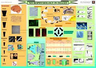
Imaging. Dr Chander Lulla Consultant interventional sonologist, Director, RIA clinic, Jaslok Hospital, Mumbai. ZONAL BLOOD FLOW Aplebaum 1995. Zone 1 -- a 2 mm thick area surrounding the hyperechoic outer layer of the endometrium Zone 2 -- the hyperechoic outer layer of the endometrium

E N D
Dr Chander Lulla Consultant interventional sonologist, Director, RIA clinic, Jaslok Hospital, Mumbai
ZONAL BLOOD FLOW Aplebaum 1995 • Zone 1 -- a 2 mm thick area surrounding the hyperechoic outer layer of the endometrium • Zone 2 -- the hyperechoic outer layer of the endometrium • Zone 3 -- the hypoechoic inner layer of the endometrium • Zone 4 -- the endometrial cavity
NT- IT-NB-FMF-MMF • 45-84mm/ 11-14 WEEKS • TA/TVS similar 5% TVS recquired • MAGNIFICATION(75% of screen) • FETAL HEAD /THORAX • MID SAGITAL SECTION • FETUS IN NEUTRAL POSITION
3D ADVANTAGES • 3D technique overcomes some limitations of conventional 2D sonography • Fast scanning time • Multiple scanning planes/Any Plane scan • C PLANE >Mullerian abnormlities, Myomas , Pregnancy, EC abn , Sono Hystero etc • Allows more detailed assessment of morphologic features • Data stored/Review possibility/Highly reproducible examination-Less operator dependent
ENDOMETRIAL CA-EC Volume Estimation GRADE of CANCER- Well differentiated -Volume lower than Moderately or Poorly Differentiated Tum. Myometrial Invasion -Volume Increased.
ADNEXIAL MASSES • 3D technique overcomes some limitations of conventional 2D sonography • Fast scanning time • Multiple scanning planes/Any Plane scan • Allows more detailed assessment of morphologic features • Data stored/Review possibility/Highly reproducible examination-Less operator dependent • Several studies have evaluated the role of 3D transvaginal sonography in assessing adnexal masses, reporting controversial results.
3D Sonohysterography • ANY PLANE POSSIBILITY • VOLUME DATA STORED-Shortening the amount of time during which the uterine cavity must remain distended. • NAVIGATION through the volume data.-Reproducibility • VOLUME of myomas can be calculated.
4D Sonohysterography • Volumes are acquired up to 16 times per second • Multiplanar views displayed and updated frequently. • Coronal plane -cavity can be observed as it is distended • Image and record dynamic events such as catheter insertion and withdrawal during distension of the lower segment and cervical canal, locations that tend to be difficult to examine
MR - Inversion Mode Visualization of hypoechoic structures which are difficult or impossible to display with conventional Ultrasound techniques in a clear inverted surface rendered display, while simultanously removing information from the surrounding tissue.
TUI – Tomographic Ultrasound Imaging • Perfect overview • More details by choosing smaller slice distances. • Results become comparable to CT and MR images, • Improves interdisciplinary cooperation. • In a difficult case, a second opinion.
MONOCHORIONIC TWINS TTTSVascular anastamosis DONOR RECEPIENT BALANCED AA SUPERFICIAL VV SUPERFICIAL AV DEEP 15% TTTS UNBALANCED Twin-twin transfusion syndrome is a result of unbalanced blood flow through vascularcommunications on the chorionic plate Untreated, the prognosisis poor, with an overall perinatal mortality of 80%
INT ILIAC EXT ILIAC FIBROID POST DIV ANT DIV UTERINE ARTEY
POST-EMBOLISATION PRE-EMBOLISATION
Elasticity Imaging The Clinical Impact
Clinical Examples • Simple cyst Note: Diagnosis based on B-mode imaging
Clinical Examples • Invasive Ductal carcinoma Note: Diagnosis based on B-mode imaging
Lesions have Unique Internal Characteristics Large Cyst with debris Fibroadenoma Note: Diagnosis based on biopsy findings Ductal Carcinoma
Virtual Touch Tissue Analysis Using ARFI • Virtual Touch Tissue Imaging • Virtual Touch Tissue Quantification