1 / 19

Pacinian Corpuscles

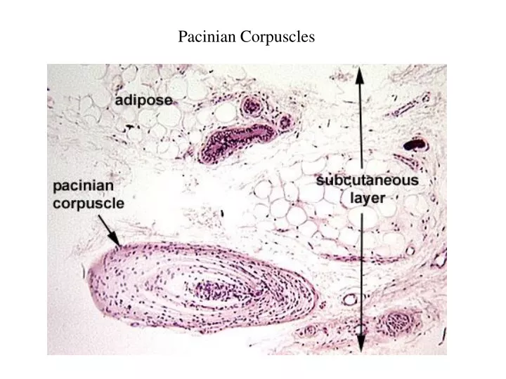
Pacinian Corpuscles. Pacinian Corpuscles. Meissner’s Corpuscles. Nasal Cavity. Nasal Cavity. Nasal Cavity. Nasal Cavity. Eye. Eye. A = Cornea B = Iris C = Retina D = Optic Nerve E = Lens F = Eyelid. Eye: Cornea. Eye: Ciliary Body. Eye: Ciliary Processes. Eye: Iris.

omerj
Download Presentation

Pacinian Corpuscles

An Image/Link below is provided (as is) to download presentation Download Policy: Content on the Website is provided to you AS IS for your information and personal use and may not be sold / licensed / shared on other websites without getting consent from its author. Content is provided to you AS IS for your information and personal use only. Download presentation by click this link. While downloading, if for some reason you are not able to download a presentation, the publisher may have deleted the file from their server. During download, if you can't get a presentation, the file might be deleted by the publisher.

E N D

Presentation Transcript


  1. Pacinian Corpuscles

  2. Pacinian Corpuscles

  3. Meissner’s Corpuscles

  4. Nasal Cavity

  5. Nasal Cavity

  6. Nasal Cavity

  7. Nasal Cavity

  8. Eye

  9. Eye A = Cornea B = Iris C = Retina D = Optic Nerve E = Lens F = Eyelid

  10. Eye: Cornea

  11. Eye: Ciliary Body

  12. Eye: Ciliary Processes

  13. Eye: Iris

  14. Eye: Lens

  15. Eye: Retina

  16. Eye: Retina

  17. Eye: Retina

  18. Choroid Plexus In the four cerebral ventricles, the ependymal layer is modified to perform a secretory function and forms the choroid plexus. In development, the innermost meningeal layer, the pia mater, in these areas becomes highly vascularized and projects into the ventricle as a series of villus-like processes called the choroid plexus (arrow). Cerebral cortex flanks the choroid plexus, and part of the cerebellum (arrowhead) is above the villi. (c) 1998 Keyboard Publishing, Inc.

  19. Choroid Plexus

More Related