Download

1 / 58
830 likes | 2.18k Views
Hydrocephalus. By M.D. Wenbing Ai. Surgical Department of Renhe Hospital of the Three Gorges University. What is Hydrocephalus ?.

E N D
Hydrocephalus By M.D. Wenbing Ai Surgical Department of Renhe Hospital of the Three Gorges University
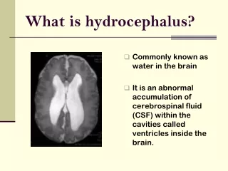
What is Hydrocephalus? • The term hydrocephalus is derived from the Greek words “hydro” meaning water and “cephalus” meaning head. As the name implies, it is a condition in which the primary characteristic is excessive accumulation of fluid in the brain. The excessive accumulation of cerebrospinal fluid (CSF) results in an abnormal dilatation of the spaces in the brain called ventricles. This dilatation causes potentially harmful pressure on the tissue of the brain.
What is Hydrocephalus? • Also can be defined: diverse group of conditions which result from impaired circulation and resorption CSF.
Pathologic specimen of brain Lateral ventricle Third ventricle Fourth ventricle
Enlarged Ventricles
Enlarged Ventricles
CSF Formation • CSF is formed by the choroid plexus . • Normal CSF production: 20 ml/h.
CSF functions CSF performs the following functions: • Balances the amount of blood in the head. • Bathes and protects the brain and spinal cord. • Carries nutrients between the brain and spinal cord while removing waste.
CSF pathway • Flows from lateral ventricles through foramina of Monro into third ventricle • Enters fourth ventricle through aqueduct of Sylvius • Enters subarachnoid space • Resorbed by arachnoid villi at top of brain
CSF homeostasis (summary) • Production: floor of the lateral ventricle and third ventricle, by choroid plexus • Circulation: L.V. -> III.V. -> IV.V. -> exits ventricular system into various basal cisterns and then to subarachroid space • Drains back to blood via arachnoid granulation to superior sagittal sinus, or via spinal nerve roots, or via olfactory tracts • Imbalance causes pathology From Johanson CE
Mechanism of hydrocephalus formation CSF * Obstruction of CSF pathway * functional disturbance of CSF absorption * Excessive production of CSF Excessive accumulation Cerebral ventricles or subarachnoid space enlargement Head enlargement Increasing ICP Cerebral dysfunction
Types of hydrocephalus According to different standards: • Noncommunicating (obstructive) hydrocephalus and communicating hydrocephalus • Congenital hydrocephalus(developed before birth) and acquired hydrocephalus(developed during or after birth) • Normal pressure hydrocephalus (NPH) and increasing pressure hydrocephalus • Acute hydrocephalus and chronic hydrocephalus
Types of hydrocephalus • Communicating hydrocephalus:the obstruction occurs in the subarachnoid space. • Noncommunicating hydrocephalus: the obstruction is located within the ventricles.
Types of hydrocephalus • Obstructive hydrocephalus • Congenital malformations • After inflammation or hemorrhage • Mass lesions • Communicating hydrocephalus • Overproduction of CSF • Defective absorption of CSF • Venous drainage insufficienc
Causes of hydrocephalus • Congenital causes in infants and children • Stenoses of the aqueduct of Sylvius due to malformation: 10% of all cases of hydrocephalus in newborns. • Dandy-Walker malformation: 2-4% of newborns with hydrocephalus. • Arnold-Chiari malformation type 1 and type 2 • Agenesis of the foramen of Monro • Congenital toxoplasmosis • Bickers-Adams syndrome: an X-linked hydrocephalus accounting for 7% of cases in males. It is characterized by stenosis of the aqueduct of Sylvius, severe mental retardation, and in 50% by an adduction-flexion deformity of the thumb.
Causes of hydrocephalus • Acquired causes in infants and children • Mass lesions account for 20% of all cases of hydrocephalus in children. • Intraventricular hemorrhage • Infections: Meningitis (especially bacterial) and cysticercosis • Increased venous sinus pressure: related to achondroplasia, some craniostenoses, or venous thrombosis. • Iatrogenic: Hypervitaminosis A, by increasing secretion of CSF or by increasing permeability of the blood-brain barrier • Idiopathic
Causes of hydrocephalus • Causes of hydrocephalus in adults • Subarachnoid hemorrhage (SAH) causes one third of these cases by blocking the arachnoid villi. • Idiopathic hydrocephalus represents one third of cases of adult hydrocephalus. • Head injury, through the same mechanism as SAH, can result in hydrocephalus. • Tumors can cause blockage anywhere along the CSF pathways. • Posterior fossa surgery • Congenital aqueductal stenosis causes hydrocephalus but may not be symptomatic until adulthood. • Meningitis, especially bacterial, may cause hydrocephalus in adults. • All causes of hydrocephalus described in infants and children are present in adults who have had congenital or childhood-acquired hydrocephalus.
Causes of hydrocephalus • Causes of NPH • SAH • Head trauma • Meningitis • Tumors • Posterior fossa surgery • Idiopathic, probably related to a deficiency of arachnoid granulations
Clinical features • Clinical features of hydrocephalus are influenced by the following: • Patient's age • Cause • Location of obstruction • Duration • Rapidity of onset
Clinical features • Symptoms in infants • Poor feeding • Irritability • Reduced activity • Vomiting
Clinical features • Symptoms in children • Slowing of mental capacity • Headaches (initially in the morning) that are more significant than in infants because of skull rigidity • Neck pain suggesting tonsillar herniation • Vomiting, more significant in the morning • Blurred vision - Consequence of papilledema and later of optic atrophy • Double vision - Related to unilateral or bilateral sixth nerve palsy • Stunted growth and sexual maturation from third ventricle dilatation: • Difficulty in walking secondary to spasticity • Drowsiness
Clinical features • Symptoms in adults • Cognitive deterioration. • Headaches: These are more prominent in the morning. As the condition progresses, headaches become severe and continuous. • Neck pain: If present, indicate herniation of cerebellar tonsil . • Nausea. • Vomiting: Sometimes explosive, more significant in the morning. • Blurred vision. • Double vision from sixth nerve palsy • Difficulty in walking • Drowsiness • Incontinence: This indicates significant destruction of frontal lobes and advanced disease.
Clinical features • Symptoms of NPH • Gait disturbance is usually the first symptom and may precede other symptoms by months or years. • Dementia presents as an impairment of recent memory or as a "slowing of thinking." The degree can vary from patient to patient. • Urinary incontinence presents as a lack of or diminished awareness of the need to urinate. Some patients may have urgency. • Other symptoms that can occur include aggressive behavior, Parkinsonlike symptoms, and seizures.
Clinical features • Sign in infants • Head enlargement. • Dysjunction of sutures. • Dilated scalp veins. • Tense fontanelle: The anterior fontanelle in infants may be excessively tense. • Setting-sun sign: • Increased limb tone.
Clinical features • Sign of children • Papilledema. • Failure of upward gaze. • Macewen sign: A "cracked pot" sound is noted on percussion of the head. • Unsteady gait. • Large head. • Unilateral or bilateral sixth nerve palsy.
Clinical features • Sign of adults • Papilledema: If raised ICP is not treated, it will lead to optic atrophy. • Failure of upward gaze and of accommodation indicates pressure on the tectal plate. • Unsteady gait is related to truncal and limb ataxia. Spasticity in legs also causes gait difficulty. • Large head: The head may have been large since childhood. • Unilateral or bilateral sixth nerve palsy is secondary to increased ICP.
Clinical features • Sign of NPH • Muscle strength is usually normal. No sensory loss is noted. • Reflexes may be increased, and Babinski response may be found in 1 or both feet. • Difficulty in walking varies from mild imbalance to inability to walk or to stand. Gait is characterized by short steps, inability to raise legs, and almost continuous activity in antigravity muscles. The patient cannot perform tandem walking and sways during Romberg test with eyes open or closed. • Sucking and grasping reflexes appear in late stages.
Diagnosis • How to diagnose hydrocephalus: • Cases history. • Symptoms and signs mentioned above. • diagnostic imaging like Skull X-rays, echoencephalogram ultrasound, CT, and MRI. • Lumbar punctures ultrasound CT MRI
Normal brain MRI imaging T1WI T1WI T1WI T2WI T2WI T2WI
Treatment of hydrocephalus • The main goal is to minimize or prevent brain damage by improving CSF flow. • Main therapy: • Medical treatment:To decrease the ICP or reduce the production of CSF with drugs. • Surgical interventions.
Surgical interventions • Treating the causes:searching for and direct removal of the obstruction is the best strategy. • Shunt procedure:the main ways are V-P shunt and V-A shunt, the most common is V-P shunt. • Third Ventriculostomy
Shunt procedure A surgical shunt within the brain may allow CSF to bypass the obstructed area, if obstruction cannot be removed.