Download
1 / 30
310 likes | 489 Views
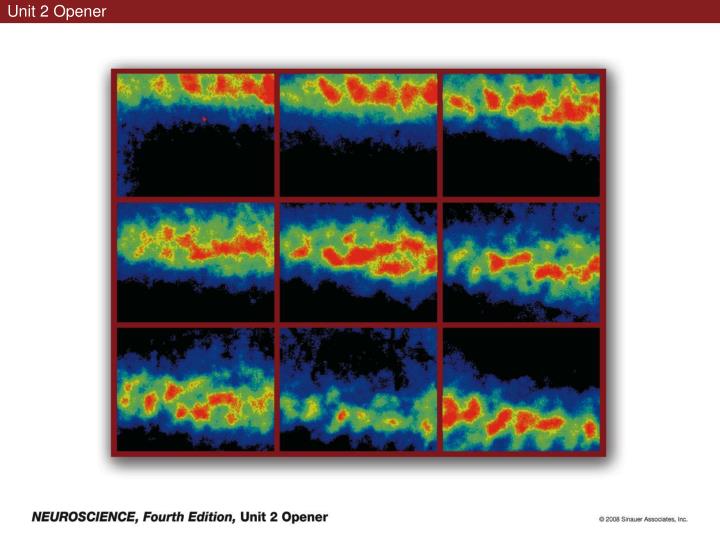
Unit 2 Opener. Figure 9.1 Somatosensory afferents convey information from the skin surface to central circuits. Figure 9.1 Somatosensory afferents convey information from skin surface to central circuits (Part 1).

E N D
Figure 9.1 Somatosensory afferents convey information from the skin surface to central circuits
Figure 9.1 Somatosensory afferents convey information from skin surface to central circuits (Part 1)
Figure 9.1 Somatosensory afferents convey information from skin surface to central circuits (Part 2)
Figure 9.2 Transduction in a mechanosensory afferent (a Pacinian corpuscle)
Figure 9.3 Receptive fields and two-point discrimination threshold
Figure 9.3 Receptive fields and two-point discrimination threshold (Part 1)
Figure 9.3 Receptive fields and two-point discrimination threshold (Part 2)
Figure 9.4 Slowly and rapidly adapting mechanoreceptors respond differently to a stimulus
Figure 9.5 The skin harbors a variety of morphologically distinct mechanoreceptors
Figure 9.6 Simulated activity patterns in different mechanosensory afferents as Braille is read
Figure 9.7 Proprioceptors provide information about the position of body parts
Figure 9.8 Schematic representation of the main mechanosensory pathways
Figure 9.8 Schematic representation of the main mechanosensory pathways (Part 1)
Figure 9.8 Schematic representation of the main mechanosensory pathways (Part 2)
Figure 9.9 Proprioceptive pathways for the upper and lower body
Figure 9.10 Somatic sensory portions of the thalamus and their cortical targets in postcentral gyrus
Figure 9.11 Somatotopic order in the human primary somatic sensory cortex
Figure 9.11 Somatotopic order in the human primary somatic sensory cortex (Part 1)
Figure 9.11 Somatotopic order in the human primary somatic sensory cortex (Part 2)
Box 9B Patterns of Organization within the Sensory Cortices: Brain Modules
Figure 9.12 Connections within the somatosensory cortex establish functional hierarchies
Figure 9.13 Neurons in the primary somatosensory cortex form functionally distinct columns
Figure 9.13 Neurons in primary somatosensory cortex form functionally distinct columns (Part 1)
Figure 9.13 Neurons in primary somatosensory cortex form functionally distinct columns (Part 2)
Figure 9.14 Changes in somatic sensory cortex of an owl monkey following amputation of a digit
Figure 9.15 Functional expansion of a cortical representation by a repetitive behavioral task