Download
1 / 15
150 likes | 159 Views
1. lL^=3?. Practical cytogenetic Dr.Tamara Sami Naji 2017-2018. 1- Cuvets 2- Syringes 3- Heparinised tubes for blood collection 4- Disposable Petri dishes. The safety in laboratory :-

E N D
1 lL^=3?
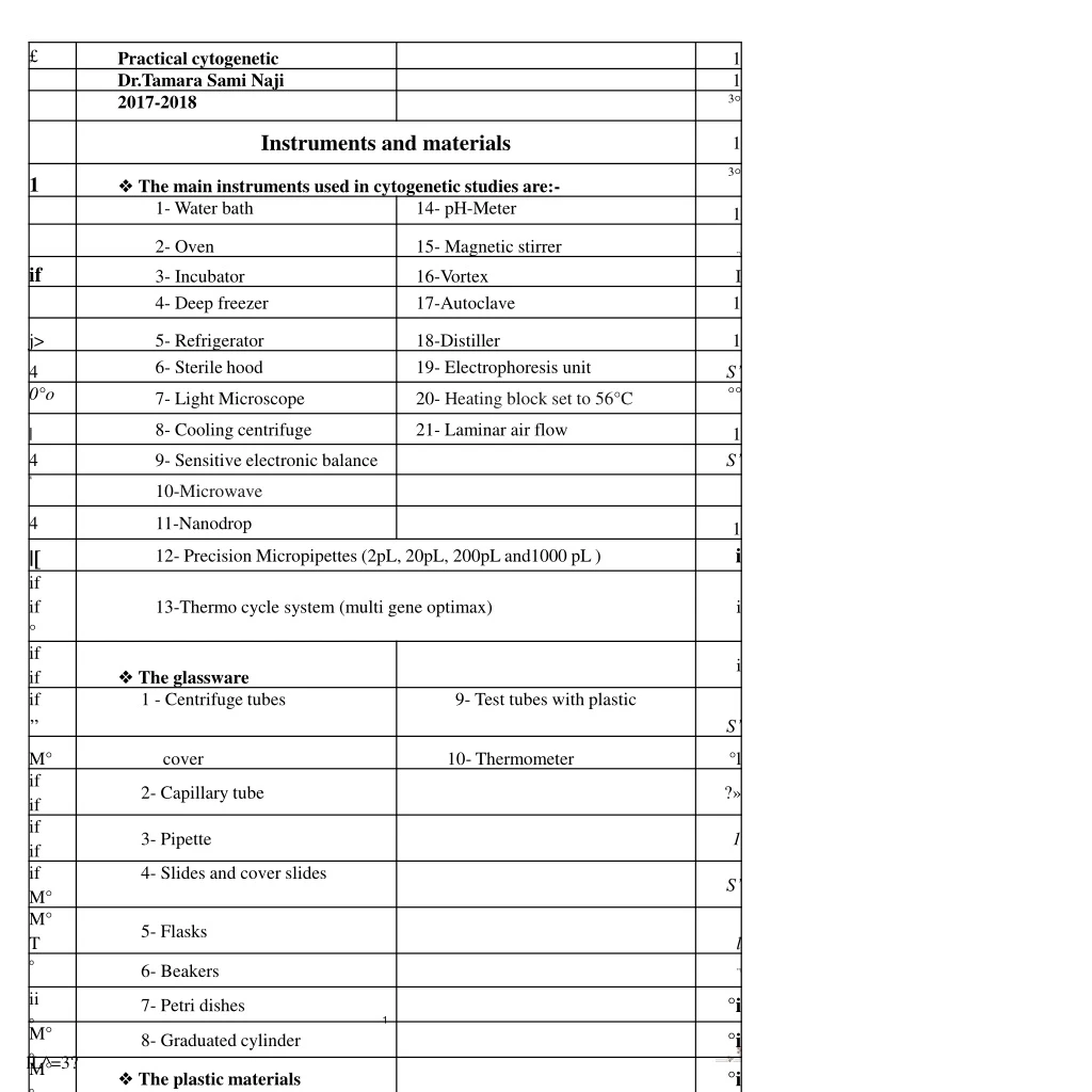
Practical cytogenetic Dr.Tamara Sami Naji 2017-2018 1- Cuvets 2- Syringes 3- Heparinised tubes for blood collection 4- Disposable Petri dishes. The safety in laboratory :- The clinical laboratory presents many hazard because of the nature of the work done there like the open flames, the glassware, chemicals of varying reactivity, flaming solvents and toxic fumes and so on. So the main rules in laboratories are:- 1- Each student must wear the laboratory coat. 2- Pipette all solutions by mechanical suction or an aspirator bulb, never use the mouth. 3- Do not use any flammable solvents and reagents near an open flame. 4- Handle all flammable solvents and reagents under a fume hood. 5- Wear gloves when handling infections substances near an open flame. 6- If a strong acids or bases spilled, wipe them up immediately using copious amount of water and great care. 7- Know where the fire extinguisher is located and how to use it properly. 8- To remove a solid reagents use a spatula. 9- Label all laboratory bottles. 10- Never return an excess reagent to its bottle. 11- Replace covers, tops or cork on all reagents bottles as soon as no longer are used. 12- Never taste any chemical and be caution when you smell a liquid. 2
Practical cytogenetic Dr.Tamara Sami Naji 2017-2018 13- Always pour acids into water for dilution never do the opposites. And pour strong acids and basis slowly to the side of the receiving vessel to prevent splashing. 14- If blood or another body fluid comes in contact with the mouth, split it out immediately into sink, rinse with mouthfuls of tap water, never swelling but discarding each mouthful. Medical genetics Medical genetics:- it involves any application of genetics to medical practice. It includes 1. Studies of the inheritance of disease in families. 2. The mapping of disease genes to specific locations on chromosomes. 3. Analysis of the molecular mechanisms through which genes cause disease. 4. The diagnosis and treatment of genetic disease. The knowledge of medical genetics is important for health care, because genetic disease make up a large proportion of the total disease burden in both paediatric and adult populations. Types of genetic disease 1. Chromosomal disorder:- when large segment or the whole chromosome is missing , duplicated or altered, these disorders include diseases such as down syndrome and turner syndrome. 2. Disorders in which single genes are altered (Mendelian) conditions or single gene disorder for example cystic fibrosis, sickle cell disease and haemophilia. 3
Practical cytogenetic Dr.Tamara Sami Naji 2017-2018 3. Multifactor disorders:- which are due to combination of multiple genetic as well as environmental causes. Many birth defect, for example cleft lip and / or cleft palate, heart disease and diabetes. 4. Mitochondrial disorders:- small number of disease caused by alterations in the small cytoplasmic mitochondrial chromosomes. Mitosis VIDEO https://www.youtube.com/watch?v=DwAFZb8juMQ&t=131s Interphase ► Prophase ► Prometaphase -► Metaphase mitotic spindle centrosomes centromeres nucleus diploid cell chromosomes (DNA) nucleus dissolves chromosomes align N=3 chromosomes replicates mitotic spindle (microtubules at center of cell attaches to centromeres Anaphase ► Telophase ► Cytokinesis mitotic spindle pulls sister mitotic spindle dissapears, cell divides, chromatids to opposite poles nuclear envelope begins two daughter cells form to reform, cell division beams Meiosis VIDEO
Practical cytogenetic Dr.Tamara Sami Naji 2017-2018 https://www.youtube.com/watch?v=nMEyeKQClqI MEIOSIS I Spindle fibers Nuceus Nuc ear envelope Prophase I (early) Prophase (late) Metaphase I Anaphase I Telophase I Synapsis and crossing Chromosomes condense Paired homologous Homologous Nuclear envelopes partially over occurs. become visible. Spindle chromosomes align chromosomes separate assemble around chromo¬ forms. Nuclear envelope along equator of cell to opposite poles of cell somes. Spindle disappears. fragments. Spindle fibers Cytokinesis divides cell into attach to each chromosome. RMO Figure 3-4 Meiosis. An actual human cell undergoing meiosis has 23 chromosome pairs. MEIOSIS II Interphase f/rM Prophase II Metaphase Anaphase II Telophase II Four nomdentical Nuclear envelope Chromosomes align along Sister chromatids separate Nuclear envelopes assemble haploid daughter to opposite poles of cell. fragments. Spindle equator of cell. around two daughter nuclei. cells forms and fibers Chromosomes decondense attach to both Spindle disappears Cytokinesis divides cells chromosomes Karyotyping 5
Practical cytogenetic Dr.Tamara Sami Naji 2017-2018 A karyotype is the number and appearance of chromosomes in the nucleus of a eukaryotic cell. The term is also used for the complete set of chromosomes in a species or in an individual organism and for a test that detects this complement or measures the number. Karyotypes describe the chromosome count of an organism and what these chromosomes look like under a light microscope. Attention is paid to their length, the position of the centromeres, banding pattern, any differences between the sex chromosomes, and any other physical characteristics. The preparation and study of karyotypes is part of cytogenetics. Karyotyping is a test to examine chromosomes in a sample of cells, which can help identify genetic problems as the cause of a disorder or disease. This test can: • Count the number of chromosomes • Look for structural changes in chromosomes How the test is performed The test can be performed on almost any tissue, including: • Amniotic fluid • Blood • Bone marrow • Tissue from the organ that develops during pregnancy to feed a growing baby (placenta) The laboratory specialist uses a microscope to examine the size, shape, and number of chromosomes in the cell sample. The stained sample is 6
Practical cytogenetic Dr.Tamara Sami Naji 2017-2018 photographed to shows the arrangement of the chromosomes. This is called a karyotype. Normal Results • Females: 44 autosomes and 2 sex chromosomes (XX), written as 46, XX • Males: 44 autosomes and 2 sex chromosomes (XY), written as 46, XY Karyotyping video https://www.youtube.com/watch?v=fctIdXqRMSk Chromosomal staining and banding techniques Conventional staining Staining procedures which provide a uniform unbanded appearance to chromosomes are referred to as solid conventional staining. This method can however be useful for studies on chromosomes breakage, gaps, deletion and ring chromosomes. A much faster procedure to obtain conventional staining involves the use of Giemsa staining technique. Banding staining Early karyotypes were useful in counting the number of the chromosomes, but structural abnormalities were often undetectable such as insertion, translocation and inversion that cannot be detected in conventional Giemsa stain. Staining technique was developed in 1970s to produce the chromosomal bands characteristic of modern karyotypes that facilitates the correct identification of individual chromosomes. The main chromosomal banding techniques: 1. Quinacrine banding (Q- banding): 7
Practical cytogenetic Dr.Tamara Sami Naji 2017-2018 Was the first staining method (1970). When chromosomes stained with quinacrine or related compounds and examined by florescence microscope each pair stains in specific pattern of bright and dim bands. Q bands were used as the reference bands for the slandered classification. 2. Giemsa banding (G- banding): In this widely used technique chromosomes are treated with trypsin, which denatures chromosomal protein, then stained with Giemsa stain. The chromosomes take up stain in pattern of dark and light staining bands (G-bands). With the dark bands corresponding to the bright Q bands. 3. Reveres banding (R- banding): If the chromosomes receive a heat pretreatment, then Giemsa staining, the resulting dark and light stained bands (R- bands) are the reveres of those produced by Q &G banding. R banding gives much the same information as Q or G banding but less widely used. 4. High resolution banding: This technique, often called prophase banding is becoming widely used in clinical genetics, in this technique the chromosomes reveal a much larger number of bands (800-1400 in all) than can be seen in metaphase preparations (about 200). High resolution banding can therefore be helpful in delineating precis breakpoints or demonstrating small alterations in chromosomes structure that would not otherwise be observed. Giemsa banding technique It is the most usual method. To obtain this stain the slides should be treated with a protease such as trypsin or in hot saline citrate. 8
Practical cytogenetic Dr.Tamara Sami Naji 2017-2018 Trypsin - Giemsa banding Materials ❖ Phosphate Buffer Saline Solution (PBS) The solution was prepared by diluting the following component in 1000 mL of distilled water (D.W) in pH (7.0) . *sodium chloride (NaCL) (8 gm) *Potassium chloride (KCL) (0.2 gm) *anhydrous sodium phosphate dibasic (Na2HPO4) (0.9 gm) *anhydrous potassium phosphate monobasic (KH2PO4) (0.2gm) ❖ Trypsin solution (0.005%) The solution was prepared by dissolve (35mg) of trypsin 1.250 (Difco) in (70 ml) of (PBS), the solution is stable for approximately one day. ❖ Giemsa stain solution The solution was prepared by dissolve 2.5ml of Giemsa in 45ml of Phosphate Buffer Saline Solution (PBS) (pH 6.8). Method 1. Incubate the slides for 20-40 seconds in trypsin solution in coplin jar. 2. Rinse the slides thoroughly with cold (refrigerated) PBS. 3. Rinse the slides in distilled water and dry with air. 4. Stain for 5 minutes in Giemsa solution. 9
o°o o°o o°o o°o o°o o° o° o° o° o° o° o° o° o° o°o o°o o°o o°o o° - o° o° o° — o° o° o° o° o° o° o° o° o°~ Practical cytogenetic Dr.Tamara Sami Naji 2017-2018 Cytogenetic X n a: c 6 c r, - O V' f H5W1 c/r tn O Tj X hi 22 21 20 19 S D 3 Variable bands B3Sj|Positive Q and G bands "■ Negative R bands □ Negative or pale-staining Q and G bands Positive R bands Fiaure 2 3 The Paris classification: a diagrammatic representation ol the human chromosomes showing Ihe bancMg poMefns and numbering scheme odopled o. me Peis commence. Keprlnled Iron, pops Conference (1971). Standardization in human cytogenetics. Birth Defects. 1972,8(7). _Op_Op__op_Op__op_Op__I r_Oq_Op_Op_Op_Op_Op_Op_Op_Op_Op_Op_Op_Op_Op_Op_Op_Op_Op_Op_Op_Op_Op_Op_Op_Op_Op_Op_0@0_]J Op_ °p__Op_Op__Op_Op__°o__gp__Op__Oq__gp__gp__gp__go__Op__gp__go__°p__°p__°p__°p__°p_°p_°p_°p_go_go_go_go_go_go_go_go_go_go_go_gp_gp_gp_Op.
Practical cytogenetic Dr.Tamara Sami Naji 2017-2018 Cytogenetic: is the study of genetic material at the cellular level. In the routine laboratory environment human cytogenetic is almost always concerned with light microscope studies of the chromosome. Blood culture Blood is one of the most accessible human tissues for chromosome analysis because growth potential after mitogen stimulation is excellent. It is also one of the easiest tissues to study because the cell cycle is well characterized. The cell can be synchronized of the preparation of long chromosomes with high resolution banding and rapid results can be obtained as sufficient mitotic figure are available for analysis after 2-3 days in culture. Blood cells involved and how can be stimulated Blood contains a number of different cell types. In normal blood neither red blood cells nor platelets contain nuclei, so that chromosomes and DNA studies are only possible on nucleated WBCs. Spontaneous division among un-stimulated WBCs either in vivo or in vitro are rare unless the individual from whom they derived had blast cells in his peripheral blood. The challenge which arises in samples for cytogenetic analysis is to induce some of white cells presents in blood sample to divide, as well known the types of WBCs in blood are • Granulocytes • Monocytes • Lymphocytes Lymphocytes (T-lymphocytes) are concerned with cell mediated immunity are of three types T- helper, T-suppressor and T-cytotoxic cells with different surfaces markers. In normal adult about 70% of 11
Practical cytogenetic Dr.Tamara Sami Naji 2017-2018 lymphocytes {1.5 - 4.0 x10⁹} per litter of blood are T-lymphocytes, the remainders are B-lymphocytes so it becomes the cell of choice. Short term blood culture depends on the ability to find cellular growth factors (mitogenes) which will induce resting or quiescent T-lymphocytes to divide by activating complex pathways which involves. ❖ T-cell antigen receptors complex ❖ Tyrosine kinesis ❖ Lymphokines There are several lymphokines but the most important one is Phytohemagglutinin PHA-stimulated response, interlukin-2. Mitogens: they are numbers of agents other than PHA, interlukin-2 or Epstein-Barr virus EBV, which will cause blood cell to become mitotically active in cell culture, by activating complex pathways which involve a number of specific intermediates, they include substances such as concavalin -A pokeweed mitogen (PWM). Preparation of chromosomes from human peripheral blood Lymphocyte blood culture ❖ The chemicals 1- The media used for blood collection (RPM1-1640, L-15, TC) 2- Antibiotic (Penicillin ,Streptomycin, Nystatin) 3- Fetal calf serum or human plasma as growth factor. 4- PHA (phytohaemaglutinin) growth factor. 5- Heparin ( anticoagulants) 6- Colsemide: It depolymerises microtubules and limits microtubule formation. 12
Practical cytogenetic Dr.Tamara Sami Naji 2017-2018 7- Hypotonic solution (KCL) for cell lyses 8- Methanol and glacial acetic acid (fixative) 9- Chemical stains(Giemsa stain) 10- Deionized distilled water 11- Buffers (NAHCO3) keep the PH at 6.8 12- Biological materials (blood, body fluid, lymph tissue like liver kidney bone marrow). ❖ Hams F10 or RPMI 1640 culture media ❖ Fetal calf serm (10%) ❖ PHA(1%) ❖ Penicillin (5000 IU/ml) (1%) ❖ Streptomycin (5000 ug/ml) (1%) ❖ L-glutamine (200 ug/ml) (1%) ❖ Colcemid (10 ug/ml) ❖ KCL (0.075 M) ❖ 3 part of methanol to 1 part glacial acetic acid Fetal calf serm: it is very important to culture media because it enriched the media with proteins that is used to cell growth and division. PHA: its phytohaemaglutinin used as mitogen to activates the lymphocytes to become mitotically active, and that used for • WBC isolation • Stimulation WBCs for growth and division The antibiotics: it is used in culture media to prevent the growth of microorganisms. 13
Practical cytogenetic Dr.Tamara Sami Naji 2017-2018 Colcemid: is added prior harvesting it acts as arresting agents because it prevent cell division in metaphase with destroys the spindle fibers during the metaphase so the two sister chromatids of the chromosome still attached and then allows us to examine the chromosomes at its contracted forms. KCL: it is a hypotonic solution used to enlarge the cell and make it easy to be ruptured and split out the chromosome inside it during the dropping. Fixative (methanol and glacial acetic acid): it is very important to fix the chromosomes and make them hard and tuff to be broken. Tissue Culture Procedure - Place (0.5) mL of heparinized blood into a culture vessel added to it (4.0) mL of culture medium (RPMI-1640) containing phytohemagglutinin (PHA) and antibiotics. - Seal the lid securely, and then Mix the contents of the vessel by gentle shaking after that incubate at 37°C ± 0.5°C in the dark for (72) h. Harvesting ❖ Add (0.1) mL of Colcemid stock solution at a concentration of(10^g/ml ) to the culture and shake gently Return to the incubator for three hours. ❖ Tip the contents of the culture vessel into a centrifuge tube. Was centrifuged at (1500) rpm for (10) min. ❖ Removed the supernatant and resuspend the pellet cells in (10) mL of 0.075M potassium chloride solution was gradually added to her and suspension was mixed thoroughly. Cells were incubated in (37°C) water bath for (30) min and inverted occasionally. 14
Practical cytogenetic Dr.Tamara Sami Naji 2017-2018 ❖ Tubes were recentrifuged at (1500) rpm for 10 min. then Removed supernatant and resuspend the cells in (10) mL of freshly prepared freezer -chilled fixative solution 3:1 (methanol / acetic acid ). The fixative must be added slowly, but at constant rate with vigorous agitation, ideally using a vortex mixer, to prevent the cell button from becoming a solid clump, and refrigerated at (4°C ) for (1) hr. ❖ Tubes were recentrifuged at (1500) rpm for 10 min. then Removed supernatant and resuspend the cells in (10) mL of fixative, This procedure repeated four times. ❖ After the last wash (1) ml of the fixative solution was added resuspended and made ready for slide preparation. 1. Slide preparation Cells suspension dropped using a Pasteur pipette , onto pre cleaned slides dipped in cold distilled water and allowed to (4-5) drops of cells suspension to drip onto the slide from a height of at least (30) cm. to assist metaphase spread . Prepare at least (4) slides from each culture. Placed the slides to dry in warm place. 15 lL^=3?
