Download
1 / 48
530 likes | 1.03k Views
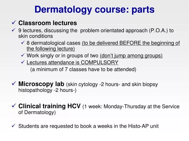
Dermatology course : parts. Classroom lectures 9 lectures, discussing the problem orientated approach (P.O.A.) to skin conditions 8 dermatological cases (to be delivered BEFORE the beginning of the following lecture) Work singly or in groups of two (don ’ t jump among groups)

E N D
Dermatology course: parts • Classroom lectures • 9 lectures, discussing the problem orientated approach (P.O.A.) to skin conditions • 8 dermatological cases (to be delivered BEFORE the beginning of the following lecture) • Work singly or in groups of two (don’t jump among groups) • Lectures attendance is COMPULSORY (a minimum of 7 classes have to be attended) • Microscopy lab (skin cytology -2 hours- and skin biopsy histopathology -2 hours-) • Clinical training HCV (1 week: Monday-Thursday at the Service of Dermatology) • Students are requested to book a weeks in the Histo-AP unit
Dermatology course: learning objectives • How to approach a dermatological case • Taking a history • Performing a physical examination/dermatological examination • Define the main problem of the patient • Make a DD and a diagnostic approach for the main dermatologic presentations: nodules, pruritus, alopecia,.. • Perform and interpret main diagnostic tests (skin scrapings, cytology, serology tests...) • Clinical management of most prevalent skin diseases of dogs and cats (aprox. 25 diseases) • Obtain and use updated medical information … you will enjoy/discover the pleasure of learning!
Dermatology course: evaluation • The final note is based on: - case work ups (40% final mark) - 2 exams (15% + 25% final mark) - evaluation of practical work at the HCV(knowledge, clinical skills, attitude,..) (20% final mark) Exams dates: October 17th 2011 (Mid term examination) November 28th 2011 (Final examination) • Type of exam: multiple choice questions
Professors • Lluís Ferrer (lectures, clinical training at the VTH) • lluis.ferrer@uab.cat • 935811421 • Mondays 11.00-13.00 at office V0-025 • Wednesdays at the VTH • Mar Bardagí (lab, clinical training at the VTH) • mar.bardagí@uab.cat • 935811421 • Wednesdays 15.00-17.00 at office V0-025 • Tuesdays at the VTH • Iván Ravera, (clinical training at the VTH) • ivan.ravera@uab.cat
Methods of Diagnosis • “CLINICAL EYE” • PERFORMING SUCCESIVE TESTS • PROBLEM ORIENTED APPROACH
“CLINICAL EYE” Diagnosis relies on a non systematic and non contrasted clinical experience Strong component of subjectivity and intuition Highly prestigious during the past, specially among people without medical education
“CLINICAL EYE” • Quick method • Cheap • If effective, generates confidence • Commonly fails • It can not be repeated and it can not be taught • It does not allow to progress Fastest way to reach a wrong diagnosis!!!
SUCCESIVE DIAGNOSTIC TESTS Different tests are performed until an alteration is found The disease is argued on the basis of the alteration found, which supposedly explains the clinical signs Apparently, the diagnosis is well-founded
SUCCESIVE TESTS II • Slow and unpredictable • Depending on the test chosen in the first place, a different diagnosis will be reached • Expensive, a lot of useless tests are performed; the owners get “tired” of it • It can not be explained and systematized easily
PROBLEM ORIENTED APPROACH The one taught nowadays in veterinary faculties: it can be explained and taught It mixes subjective decisions (problem definition) with science based actions It is rather save and effective Minimum expenses to reach the diagnosis
How to make a diagnosis (I) Signalment, chief complain, history Physical and dermatological examination Problem
How to make a diagnosis (II) Problem Differential Diagnosis Diagnostic Tests Diagnosis
Signalment • Species • Age • Breed (coat color) • Sex (neutered or not)
Chief complaint cause and duration of the owner’s concern
Dermatological and medical history • Dermatological history: • age at onset • original lesion’s location and initial appearance • progression of the lesions • pruritus (grade) • response to treatment/s • contagion to other animals/people • Medical history • habitat, diet, other signs,...
General physical examination and dermatological examination • General physical examination • Dermatological examination: • visual and “manual” examination • all the skin surface (included external ear canals) • presence of parasites • skin and hair quality • type of cutaneous lesions • distribution of the lesions
Skin and hair quality • Skin: • Color • Thickness • Elasticity • Hair: • Texture (broken, oily, dry…) • Easy to epilate • Primary/secondary hairs ratio
Fundamental cutaneous lesions • Primary • Secondary (to self-trauma and/or to spontaneous progression of primary lesions)
Primary cutaneous lesions • Macule or patch (màcula) • Papule (pàpula) • Plaque (placa) • Pustule (pústula) • Vesicle or bulla (vesícula o bulla) • Wheal (fava) • Nodule (nòdul) • Tumour (tumor)
Macule • Circumscribed area, up to 1 cm in diameter, characterized by a change in the color of the skin • erythematous • hyperpigmented (melanotic) • Haemorrhagic • Patch: diameter > 1 cm
Papule • Small solid elevation of the skin up to 1 cm in diameter • Normal color, erythematous, hyperpigmented • Plaque:from coalescing papules (diameter > 1 cm)
Pustule • Small elevation of the skin filled with pus • Fragile! • Follicular / Non-follicular
Wheal • Sharply circumscribed, raised lesion (dermal edema) • Usually erythematous • Variable shape and size • Transient
Nodule/tumour • Solid elevation greater than 1 cm in diameter • Variable depth and attachment to underlying tissues • Inflammatory/neoplastic (!!)
Erythema (not a “pure” cutaneousprimary lesion) • Change in the color of the skin (“redness”) • Variable limits,shape, size • Blanch on diascopy ( with hemorrhage)
Cutaneous lesions that may be primary or secondary • Alopecia (alopècia) • Scale (escates) • Follicular cast (cilindre fol.licular) • Crust (crosta) • Comedo (comedó) • Pigmentary abnormalities
Alopecia • Lack of hair where it is normally present • Complete alopecia • Partial alopecia (hypotrichosis): due to reduction of number and/or length of hairs
Scale • Loose fragment of stratum corneum • Variable size, color, consistency • Follicular cast
Crust • Accumulation of dried exudate, blood, cells (epithelial and inflammatory), hairs, adhered to the skin. • The underlying skin is excoriated/eroded/ ulcerated • Variable color
Comedo • Dilated hair follicles, which appear full of keratinaceous material • Clinically appear as “black points”
Hyperpigmentation/Despigmentation • Increased pigmentation (melanin) • of the skin • Variable distribution, shape, size • With/without inflammatory changes of the skin
Secondary cutaneous lesions • Epidermal collarette • Excoriation (escoriació) • Erosion (erosió) • Ulceració (ulceració) • Lichenification (liquenificació)
Erosion/Excoriation/Ulcer • Break in the continuity of the skin with exposure of the dermis • Variable depth, shape, bleeding • Erosion: more superficial defect without damage of the basal membrane • Excoriation: self-produced erosion, may be linear in configuration
Lichenification • Thickening of the skin characterized by exaggerated skin markings (“wrinkles”) • Usually due to chronic trauma (pruritus) • More frequent in the ventral skin • Often accompanied by hyperpigmentation
How to make a diagnosis (I) Signalment, chief complain, history Physical and dermatological examination Problem
Definition of the problem/s (P.O.A) • Definition of the problem/s is based on: • signalment, history (i.e. pruritus) • dermatological examination (“distinctive” lesions and their distribution - i.e. symmetric alopecia) • The definition of the problem is diagnosis-oriented
How to define the problem • It is the most important, distinctive and useful clinical-dermatological pattern • It is a subjective decision, based upon experience and knowledge • It is possible to define more than one problem in one patient
Differential diagnosis (DDX) • includes 3-5 diseases (or group of diseases) in decreasing order of probability • is based on the signalment and the problem/s identified during the examination
How to make a diagnosis (II) Problem Differential Diagnosis Diagnostic Tests Diagnosis
Diagnostic procedures/tests • Skin scrapings, microscopic examination of plucked hairs, cytological examination • Wood’s lamp, fungal culture, scotch test • Bacterial culture and sensitivity test • Elimination diet, allergy testing • Skin biopsy • CBC, biochemistry profile and urinalysis, serologic tests, endocrine function tests
Comments • The diagnostic procedures have to be performed in a logical sequence • Directly related to the diseases included in the differential diagnosis • The objective of the dermatological examination is the identification of the cause/s of the disease (definitive diagnosis)
EVIDENCE BASED DERMATOLOGY (EBD)the future that comes “conscientious, explicit and judicious use of currentbest evidence in making decisions about the care of individual patients” Diagnosis, treatment and prognosis are based on published scientific clinical data, not upon clinical experience, “authorities” opinions or internet….
EBD in practice Based upon traditional clinical practice and bibliography search and study • Pose a question related to the patient • Find out the answer by searching into scientific information • Be critical with the information obtained • Apply the information to the patient
EBD: advantages • It offers the best option to the patient (it is the kind of medicine that everybody would like to receive…) • It is well systemized and explained • It helps to the clinician formation in the everyday clinical practice • Protects the clinician (it can be proven that what was done was the best…!)
EBD: difficulties • Good medical formation is required • It requires good scientific information • It requires time • In veterinary medicine, good scientific evidence is scarce
EBD: which is the best evidence? • Sytematic reviews and meta-analysis of randomized trials • RCT (Randomized Controlled Trials) • Non-randomized study • No controlled study (case series) • Case report (published) • Anecdote
Dermatological case • Definition of the problem/s • concise • do not repeat the description of the lesions! • Differential diagnosis (DDX) • include only the diseases (not too many!) which can cause the lesions/signs described • don’t explain (copy…) the clinical features of the diseases
Dermatological case • Diagnostic plan • case-oriented (not problem-oriented!) • consider only diagnostic procedures useful to rule out/confirm the diseases included in the DDX