Download
1 / 33
330 likes | 535 Views
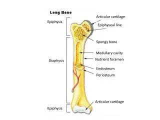
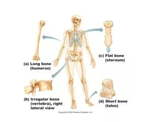
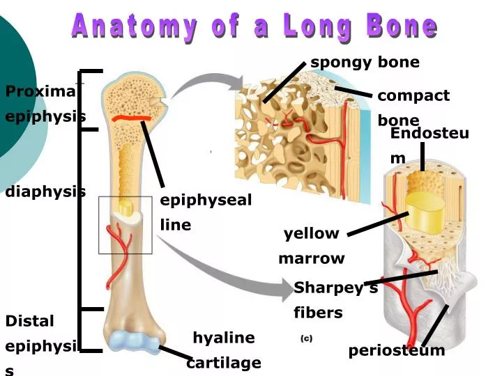
Anatomy of a Long Bone. spongy bone. Proximal epiphysis. compact bone. Endosteum. diaphysis. epiphyseal line. yellow marrow. Sharpey’s fibers. Distal epiphysis. hyaline cartilage. periosteum. Microscopic Bone Anatomy. Axial Division. Appendicular Skeleton.

E N D
Anatomy of a Long Bone spongy bone Proximal epiphysis compact bone Endosteum diaphysis epiphyseal line yellow marrow Sharpey’s fibers Distal epiphysis hyaline cartilage periosteum
Appendicular Skeleton • The Appendicular Division consists of 126 bones making up the appendages and girdles that connect appendages to the body. The pectoral girdle consists of the clavicle and scapula. The pelvic girdle is formed by the two coxal bones which are fused anteriorly.
Sphenoid Bone • The feature called the "temple" is actually a wing of the Sphenoid bone e. Sphenoid Bone – butterfly shaped bone that spans the width of the skull and forms the floor of the cranial cavity. Contains a small depression called the sella turcica (Turk’s saddle) which holds the pituitary gland in place Posterior view
Ethmoid Bone f. Ethmoid Bone – forms the roof of the nasal cavity and medial walls of the orbits
Hyoid Bone 3. Hyoid Bone – the only bone of the body that does not articulate directly with another bone, suspended in the midneck region above the larynx where ligaments anchor it to the styloid processes of the temporal bone. Serves as a movable base for the tongue and attachment for neck muscles.
Fetal Skull 4. Fetal Skull – bones of the fetal skull are not fused but contain fibrous membranes called fontanels, “soft spots” that connect the cranial bones. They allow room for the brain to grow and allow the fetal skull to be compressed during child birth. Anterior fontanel, posterior fontanel, sphenoid fontanel, mastoid fontanel
Cervical Vertebrae • Cervical Vertebrae – 7, the first two are called the atlas and axis –they allow you to rotate your head from side to side
Thoracic and Lumbar Vertebrae • 2. Thoracic Vertebrae - 12, larger than the cervical vertebrae • 3. Lumbar Vertebrae – 5, largest and sturdiest of the vertebrae Lumbar 1-Vertebral Body 2-Spinous Process 3-Transverse Facet 4-Pedicle 5-Foramen 6-Lamina 7-Superior Facet
Inferior Vertebral Column • 4. Sacrum - fuses with the coccyx inferiorly • 5. Coccyx - considered the human “tailbone”
APPENDICULAR SKELETON (pp. 138-145) A. Bones of the Shoulder Girdle, also called the Pectoral Girdle (p. 138) 1. Clavical (Collarbones) – attaches to the manubrium of the sternum medially and the scapula laterally 2. Scapulae (Shoulder Blades) – also called “wings” due to flaring when we move our arms posteriorl • Important Bone markings: • Acromion – enlarged end of the spine • Coracoid process – connects with the clavicle laterally at the acromioclavicular joint • Glenoid cavity – a shallow socket that receives the head of the humerus (arm bone)
Bones of the Upper Limb 1. Arm A. Humerus B. Important Bone Markings: Greater and lesser tubercles – sites of muscle attachments near the head of the humerus Deltoid Tuberosity – a roughened area of the shaft where the deltoid muscle attaches Trochlea – medial distal end that looks like a spool Capitulum – lateral distal end, looks like a ball Coronoid Fossa – depression above the trochlea Olecranon Fossa- posterior distal surface these two fossa’s allow the ulna to move freely when the elbow is bent and extended
Forearm Bones • Forearm • Radius – the lateral boneImportant Markings: Radial tuberosity - just below the head, where the biceps tendon attaches • Ulna - the medial bone Coronoid process- on proximal end Olecranon process – on proximal end (forms the elbow by articulating into the olecranon fossa on the humerus • Trochlear Notch – groove that separates the coronoid and olecranon processes • Interosseous Membrane- the membrane that connects the radius and ulna along its length
Hand • 3. Hand • A. Carpals – wrist bones, the most commonly fractured when falling on an outstretched hand is the scaphoid bone ( sits on thumbside) • B. Metacarpals – articulate with carpals and phalanges • C. Phalanges – finger bones
Bones of the Pelvic Girdle • Bones of the Pelvic Girdle (pp. 141-143) –formed by the two coxal bones • Coxal Bones (Hip Bones) Formed by the fusion of three bones: • Ilium – connects with the sacrum at the sacroiliac joint, large flaring bone that forms most of the hip bone. Iliac crest- upper edge
Ischium Ischium – the “sitdown” bone, it is most inferior • Ischial tuberosity - part you’re sitting on • Ischial spine - superior to the tuberosity, narrows the outlet of the pelvis Greater Sciatic notch – allows blood vessels and the large sciatic nerve to pass through
Pubis Bone Pubis – most anterior Obturator Foramen – opening which allows blood vessels and nerve to pass through to the thigh Pubic Symphysis – where the two pubic bones fuse anteriorly to form a cartilaginous joint • Acetabulum – where the ilium, ischium, and pubis fuse, receives the head of the femur
Lower Limb Bones • Bones of the Lower Limbs (pp. 143-145) • Thigh • Femur – heaviest, strongest bone in the body Greater and lesser trochanters – blunt processes at the proximal end that are sites of muscle attachments Medial and Lateral condyles – at distal end where the femur articulates with the tibia
Lower Leg Bones • Leg • Tibia – shinbone, medial weight-bearing bone of lower leg Tibial Tuberosity – proximal end of tibia where patellar ligaments and tendon attach Medial malleolus – distal inner bulge of the ankle • Fibula – stick-like lateral bone of lower leg, joins with the tibia proximally and distally Lateral malleolus – distal fibula that forms the outer part of the ankle
Foot Bones • Foot • Tarsals – 7 bones cuneiform bones (medial, intermediate, lateral) cuboid, navicular, talus, calcaneus • Metatarsals – form the sole • Phalanges - toes
Helpful Websites for Studying • http://www.gwc.maricopa.edu/class/bio201/skull/antskul.htm • http://www.getbodysmart.com/index.htm • http://www.getbodysmart.com/ap/skeletalsystem/skeleton/menu/animation.html
Q of Week: Can people grow extra bones? • multiple hereditary exostosis causes excess bones to grow in the body • Acromegaly- a disease associated with excessive bone growth • Paget's Disease • Melorheostosis-rare and progressive disorder that involves hyperostosis or thickening of cortical bone