Download
1 / 11
110 likes | 203 Views
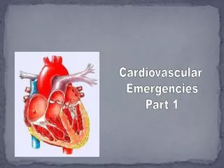
Cardiovascular Emergencies. Heart must have continuous supply of nutrients and oxygen. During exertion, myocardium requires increased supply. This is accomplished by dilation of coronary arteries. Cardiovascular System. Heart.

E N D
Cardiovascular Emergencies
Heart must have continuous supply of nutrients and oxygen. During exertion, myocardium requires increased supply. This is accomplished by dilation of coronary arteries.
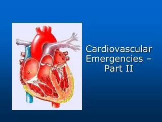
Cardiovascular System Heart
Chest pain or discomfort comes fromischemia – insufficient oxygen due to partialor complete blockage of coronary arteries.th This is most often due to atherosclerosis – buildup of plaque inside the vessel walls.
Angina Pectoris= ischemic cardiac eventin which S/S come about with exertion, but go away with rest, O2, and/or NTG Acute Myocardial Infarction = occlusive cardiac event in which S/S may occur without warning, and do not go away with rest, O2, and/or NTG. Pain of AMI differs from angina only in that -- 1. can occur at any time, without exertion 2. does not resolve in a few minutes 3. may or may be relieved by rest or NTG
What do you see - Early Cardiac Compromise Chief Complaint – Chest Pain (pressure), Dyspnea, Brain – ALOC, anxiety, feeling of Impending Doom Heart – Generally faster HR, Irregularity common (due to arrhythmia), BP normal or high Lungs – Generally normal to rapid rate, and normal to shallow TV Skin Signs – ashen gray, clammy, cool Other – nausea, vomiting, radiating pain Use OPQRST to get details of pain SAMPLE for medical history
What do you see - Late Cardiac Compromise Brain – Semiconscious to Unresponsive Heart – HR is rapid, thready, irregular BP is falling (<90) Lungs – Severe respiratory distress Rapid, shallow respirations “wet” lung sounds (fluid in lungs) Skin Signs – cyanotic, diaphoretic, cool Other – nausea, vomiting, cool to the touch Person in Decompensated Cardiogenic Shock.
Treatment 1. Put patient in position of comfort (easiest to breath) 2. Administer hi-flow O2 3. Assist ventilations if needed (PPV wi BVM) 4. Help to Administer Nitroglycerin 5. Keep patient warm 6. Prompt Transportation to EMS
Administering Nitroglycerin 1. Check medication and expiration date 2. Check there is radial pulse (want dia BP > 100) Check to make sure no ED med within 24 hours 3. Ask patient to lift tongue and have them place tablet or spray underneath tongue; keep mouth closed until dissolved 4. Watch out for fainting
But Cardiac Event is not only cause ofsimilar S&S = chest pain & dyspnea ** Treat all patients with chest pain & dyspnea as cardiac event until determined otherwise Pulmonary Embolism – blockage of pulmonary artery S&S – sudden onset of S&S; cough up blood Pericarditis – infection in pericardial sac S&S – skin in warm, feels sick & weak
Pericardial Tamponade – sac around heart fills with blood, not allowing heart to beat; result of chest trauma; S&S [Beck’s Triad] = muffled heart sounds, JVD, little difference in BP sys/dia Congestive Heart Failure (CHF)- heart muscle so damaged that cannot keep up with return flow of blood S&S – Right side = JVD, lower ext swelling Left side = pulmonary edema (pink sputum)