Download
1 / 40
510 likes | 1.09k Views
Riboswitches. Sharon Epstein 30/03/2006 Frontiers in Metabolome sciences Feinberg Graduate School. Outline. Introduction Concepts Evolution Structure Mechanisms Methods Examples Applications Conclusion. Introduction. Pubmed search for “ riboswitches ” : Reviews: 12 Articles: 57

E N D
Riboswitches Sharon Epstein 30/03/2006 Frontiers in Metabolome sciences Feinberg Graduate School
Outline • Introduction • Concepts • Evolution • Structure • Mechanisms • Methods • Examples • Applications • Conclusion
Introduction • Pubmed search for “riboswitches”: • Reviews: 12 • Articles: 57 • Pubmed search for “micro RNA”: • Reviews: 167 • Articles: 740
Introduction • Riboswitches were “discovered” in the beginning of the 21st century • The idea was known but could not be proved
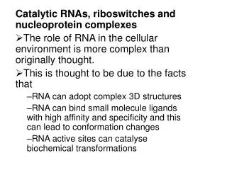
Concept • Riboswitches: “ method of controlling gene expression. (…)a sequence of RNA that, through its secondary and tertiary structure, selectively binds a specific metabolite.” (Templeton et al, 2005)
Concept • When a metabolite is bound the secondary and tertiary structure of the RNA changes affecting transcription and translation in prokaryotes and possibly mRNA processing in eukaryotes
Evolution • RNA world, possible mode of regulation in the absence of proteins • General conservation of metabolites so far known to be involved in binding • Present and studied in prokaryotes with differences in bacterial groups • Present in prokaryotes mainly on 5’UTR
Evolution • Another hypothesis: it is more recent then RNA world but is present in different bacterial groups because of lateral transfer and repetitive re-invention • Focus prokaryote
Evolution • Major difference in eukaryotes: localization • Present in introns and 3’UTR, not well studied and not much data available • Found in Arabidopsis and rice, on different splice variants (one regulated one not)
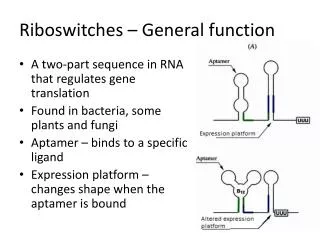
Structure • Riboswiches are composed of two interdependent but distinct domains: • Aptamer domain (responsible for binding of ligand) • Expression plataform (responsible for impacting gene expression)
Mechanism • Three known mechanisms: • Formation of intrinsic terminator stem (inhibits transcription by inducing its termination) • Formation of complex hiding translation initiation site • Self-cleaving mechanism
Methods • In line probing:
Methods • Equilibrium dialysis – radio labeled metabolite – unequal distribution • RNase H probing – DNA complementary strand – conformational change – no cleavage • Fluorescence – FMN quenched in contact with riboswitch
Coenzyme B12 • One of the first to be discovered • Upstream of cobalamin synthesis, porphyrin and cobalt transport and glutamate fermentation • One of the largest aptamers with many connecting points
Thiamine pyrophosphate • Most widespread (also found by sequence similarity in plants) • Identification of the riboswitch lead to function characterization of genes involved in the pathway
Flavin Mononucleotide • Present upstream of genes for riboflavin biosynthesis and transport pathway • Binds FMN 100 folds more tightly then riboflavin (difference: one phosphate)
Guanine and adenine • Same aptamer binds both – only one point mutation C to U (forms base-pairing with ligand) • Tertiary structure is similar – sequence only 59%
S-adenosylmethionine • S-box motif – present mainly in gram-positive bacteria • Upstream of sulfur, cysteine, SAM and methionine pathways • 1:1 stoichiomestry, dependent on Mg+2
Lysine • Descriminates between l and d lysine • AEC (toxic analog) is also bound by riboswitch and resistant bacteria carry mutations • Potential drug target?
Glucosamine-6-phosphate • RNA undergoes rapid self cleavage upon binding of metabolite • Mutations that affect ribozyme activity de-repress the gene
Glycine • Two aptamer upstream of glycine cleaving proteins – each binds one glycine but increases affinity of the other aptamer – increase in sensitivity • Evolutionary advantage?
Applications • Drug target (antimicrobial) • Molecular engineering
Conclusion • Evolutionary clues • Non coding regions of RNA as undiscovered regulatory domains • New role of bioinformatics • Possibilities
Articles • Winkler WC. (2005) • Winkler WC et al. (2005) • Tucker BJ et al. (2005) • Templeton GW et al. (2004) • Soukup JK et al. (2004) • Mandal M et al. (2004) • Nudler E et al. (2004) • Vitreschak AG et al. (2004) • Kaempfer R. (2003) • Lai EC. (2003) • Corbino KA et al. (2005) • Altman S et al. (2005)