Download
1 / 43
460 likes | 605 Views
UTERINE FIBROIDS. Dr sajda al rubai CONSULTANT OBSTETRICIAN GYNECOLOGIST. LEIOMYOMA. What is a leiomyoma? It is a benign neoplasm of the muscular wall of the uterus composed primarily of smooth muscle What is the incidence of leiomyomas? They are the most common pelvic tumors

E N D
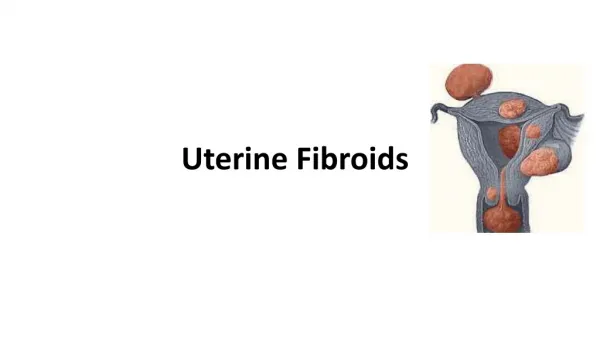
UTERINE FIBROIDS Dr sajda al rubai CONSULTANT OBSTETRICIAN GYNECOLOGIST
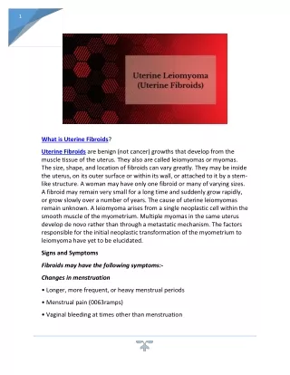
LEIOMYOMA What is a leiomyoma? It is a benign neoplasm of the muscular wall of the uterus composed primarily of smooth muscle What is the incidence of leiomyomas? They are the most common pelvic tumors It is found in 25% of white women & 50% of black women
ETIOLOGY • Unknown • Each individual myoma is unicellular in origin • Estogens no evidence that it is a causative factor , it has been implicated in growth of myomas • Myomas contain estrogen receptors in higher concentration than surrounding myometrium • Myomas may increase in size with estrogen therapy & in pregnancy & decrease after menopause • They are not detectable before puberty • Progestrone increase mitotic activity & reduce apoptosis in size • There may be genetic predisposition
PATHOLOGY • Frequently multiple • May reach 15 cm in size or larger • Firm • Spherical or irregularly lobulated • Have a false capsule • Can be easily enucleated from surrounding myometrium
CLASSIFICATION • Submucous leiomyoma • Pedunculated submucous • Intramural or interstitial • Subserous or subperitoneal • Pedunculated abdominal • Parasitic • Intraligmentary • Cervical
MICROSCOPIC STRUCTURE • Whorled appearance nonstriated muscle fibers arranged in bundles running in different directions • Individual cells are spindle shaped uniform • Varying amount of connective tissue are interlaced between muscle fibers • Pseudocapsule of areolar tissue & compressed myometrium • Arteries are less dense than myometrium & do not have a regular pattern of distribution • 1-2 major vesseles are found at the base or pedicle
1-BENIGN DEGENERATION • Atrophic • Hyaline yellow, soft gelatinous areas • Cystic liquefaction follows extreme hyalinization • Calcific circulatory deprivation precipitation of ca carbonate & phosphate • Septic circulatory deprivation necrosis infection • Myxomatous (fatty) uncommon, follows hyaline or cystic degenration
1-BENIGN DEGENRATION (cont’d) Red (carneous) degeneration • Commonly occurs during pregnancy • Edema & hypertrophy impede blood supply aseptic degenration & infarction with venous thrombosis & hemorrhage • Painful but self-limiting • May result in preterm labor & rarely DIC 2-MALIGNANT TRANSFORMATION • Transformation to leiomyosarcomas occurs in 0.1-0.5%
1-SYMPTOMS • Symptomatic in only 35-50% of Pt • Symptoms depend on location, size, changes & pregnancy status 1-Abnormal uterine bleeding • The most common 30% • Heavy / prolonged bleeding (menorrhagia) iron deficiency anemia
1-Abnormal uterine bleeding (cont’d) • Submucous myoma produce the most pronounced symptoms of menorrhagia, pre & post-menstrual spotting • Bleeding is due to interruption of blood supply to the endometrium, distortion & congestion of surrounding vessels or ulceration of the overlying endometrium • Pedunculated submucous areas of venouse thrombosis & necrosis on the surface intermenstrtual bleeding
2-PAIN • Vascular occlusion necrosis, infection • Torsion of a pedunculated fibroid acute pain • Myometrial contractions to expel the myoma • Red degenration acute pain • Heaviness fullness in the pelvic area • Feeling a mass • If the tumor gets impacted in the pelvis pressure on nerves back pain radiating to the lower extremities • Dysparunea if it is protruding to vagina
3-PRESSURE EFFECTS • If large may distort or obstruct other organs like ureters, bladder or rectum urinary symptoms, hydroureter, constipation, pelvic venous congestion & LL edema • Rarely a posterior fundal tumor extreme retroflexion of the uterus distorting the bladder base urinary retention • Parasitic tumor may cause bowel obstruction • Cervical tumors serosanguineous vaginal discharge, bleeding, dyspareunia or infertility
4-INFERTILITY • The relationship is uncertain • 27-40% of women with multiple fibroids are infertile but other causes of infertility are present • Endocavitary tumors affect fertility more 5- SPONTANEOUS ABORTIONS • ~2X N incidence before myomectomy 40% after myomectomy 20% • More with intracavitary tumors
EXAMINTION • Most myoma are discovered on routine bimanual pelvic exam or abdominal examination • Retroflexed retroverted uterus obscure the palpation of myomas LABORATORY FINDINGS • Anemia • Depletion of iron reserve • Rarely erythrocytosis pressure on the ureters back pressure on the kidneys erythropoietin • Acute degeneration & infection ESR, leucocytosis, & fever
IMAGING • Pelvic U/S is very helpful in confirming the Dx & excluding pregnancy / Particularly in obese Pt • Saline hysterosonography can identify submucous myoma that may be missed on U/S • HSG will show intrauterine leiomyoma • MRI highly accurate in delineating the size, location & no. of myomas , but not always necessary • IVP will show ureteral dilatation or deviation & urinary anomalies HYSTROSCOPY for identification & removal of submucous myomas
DIFFERENTIAL DIAGNOSIS • Usually easily diagnosed • Exclude pregnancy • Exclude other pelvic masses -Ovarian Ca -Tubo-ovarian abscess -Endometriosis -Adenexa, omentum or bowel adherent to the uterus • Exclude other causes of uterine enlargement: -Adenomyosis -Myometrial hypertrophy -Congenital anomalies -Endometrial Ca
DIFFERENTIAL DIAGNOSIS Exclude other causes of abnormal bleeding • Endometrial hyperplasia • Endometrial or tubal Ca • Uterine sarcoma • Ovarian Ca • Polyps • Adenomyosis • DUB • Endometriosis • Exogenouse estrogens Endometrial biopsy or D&C is essential in the evaluation of abnormal bleeding to exclude endometrial Ca
1-COMPLICATIONS IN PREGNANCY • ≥ 2/3 of women with fibroids & unexplained infertility conceive after myomectomy Red degeneration • In the 2nd or 3rd trimester of pregnancy rapid in size vascular deprivation degeneration • Causes pain & tenderness • May initiate preterm labor • Managed conservatively with bedrest & narcotics + tocolytics if indicated • After the acute phase pregnancy will continue to term
COMPLICATIONS IN PREGNANCY DURING LABOR • Uterine inertia • Malpresentation • Obstruction of the birth canal • Cervical or isthmeic myoma necessitate CS • PPH
COMPLICATIONS IN NONPREGNANT WOMEN • Heavy bleeding with anemia is the most common • Urinary or bowel obstruction from large parasitic myoma is much less common • Malignant transformation is rare • Ureteral injury or ligation is a recognized complication of surgery for Cx myoma • No evidence that COCP the size of myomas • Postmenopausal women on HRT must be followed up with pelvic exam or U/S every 6 M
TREATMENT DEPENDS ON: • Age • Parity • Pregnancy status • Desire for future pregnancy • General health • Symptoms • Size • Location
A-EMERGENCY MEASURES • Blood transfusion/ PRBC to correct anemia • Emergrncy surgery indicatd for: - infected myoma -acute torsion -intestinal obstruction • Myomectomy is contraindicated during pregnancy
B-SPECIFIC MEASURES • Most cases asymptomatic no treatment • Postmenopausal no treatment • Other causes of pelvic mass must be excluded • The Dx must be certain • Initial follow up every 6 M to determine the rate of growth of the myoma • Surgery is contraindicated in pregnancy • The only indication for myomectomy in pregnancy is torsion of a pedunculated fibroid • Myomectomy is not recommended during CS • Pregnant women with previous multiple myomectomy / especially if the cavity was entered should be delivered by CS to risk of scar rupture in labor
GNRH AGONISTS RX results in: 1- size of the myomas 50% maximum 2- This shrinkage is achieved in 3M of RX 3-Amenorrhea & hypoestrogenic side-effects occur 4-Osteopososis may occur if Rx last > 6M It is indicated for 1- bleeding from myoma except for the polypoid submucous type 2-Preoperative to size allow for vaginal hysterectomy myomectomy laparoscopic myomectomy
C-SUPPORTIVE MEASURES • PAP smear & endometrial sampling for all Pt with irregular bleeding • Before surgery -Correct Hb -Prophylactic antibiotics -Mechanical & antibiotic bowel preparation if difficult surgery is anticipated • Prophylactic heparin postoperative
D-SURGICAL MEASURES 1-Evaluation for other neoplasia 2-Myomectomy • For symptomatic Pt who wish to preserve fertility • Open myomectomy • Laparoscopic myomectomy • Hysteroscopic myomectomy 3-Hysterectomy • Vaginal hysterectomy • Abdominal hysterectomy 4-Uterine artery embolisation
Potential problems with hysterectomy • Post hysterectomy depression (?) • Lack of interest in sex (7%) (not significant) • Lack of enjoyment of sex (1%)
Uterine Fibroids • UAE is a fundamentally different treatment for fibroids • Minimally invasive • Low complication rates • Effective • Repeatable if necessary • Uses proved technique of embolization, available for more than 30 years
Uterine Artery Embolization • Uterine artery embolization (UAE) as a primary form of therapy was reported by Ravina in 1995 • 16 patients were treated • Polyvinyl alcohol particles used as embolic agent • Mean FU 20 months • Symptoms resolved in 11 of 16 patients • 3 partially improved • 2 failures (1 hysterectomy @ 6 weeks, 1 myomectomy @ 6 months Ravina et al. Lancet 1995
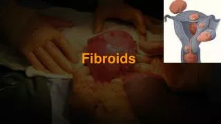
Fibroids die (caseous necrosis) • Then scar down (hyaline sclerosis) and shrink
Uterine Artery Embolization • Severe bleeding was controlled, even in women with large uteri and minimal initial volume reductions • UAE reduced the average menstrual duration from 7.6 to 5.4 days • 17 pregnancies were observed • Major complications occurred in 1% of the cases Pron et al Fertility & Sterility, 2003