Download
1 / 63
630 likes | 663 Views
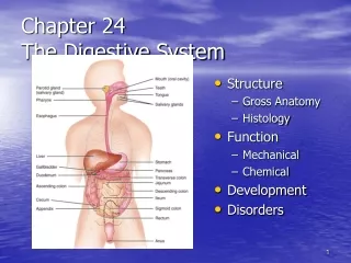
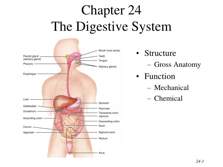
Chapter 24 The Digestive System. Structure Gross Anatomy Function Mechanical Chemical. Overview of GI tract Functions. Mouth---bite, chew, swallow Pharynx and esophagus----transport Stomach----mechanical disruption; absorption of water & alcohol

E N D
Chapter 24The Digestive System • Structure • Gross Anatomy • Function • Mechanical • Chemical
Overview of GI tract Functions • Mouth---bite, chew, swallow • Pharynx and esophagus----transport • Stomach----mechanical disruption; absorption of water & alcohol • Small intestine--chemical & mechanical digestion & absorption • Large intestine----absorb electrolytes & vitamins (B and K) • Rectum and anus---defecation
Layers of the GI Tract 1. Mucosal layer 2. Submucosal layer 3. Muscularis layer 4. Serosa layer
Mucosa • Epithelium • stratified squamous(in mouth,esophagus & anus) = tough • simple columnar in the rest • secretes enzymes and absorbs nutrients • specialized cells (goblet) secrete mucous onto cell surfaces • enteroendocrine cells---secrete hormones controlling organ function • Lamina propria • thin layer of loose connective tissue • contains BV and lymphatic tissue • Muscularis mucosae---thin layer of smooth muscle • causes folds to form in mucosal layer • increases local movements increasing absorption with exposure to “new” nutrients
Submucosa • Loose connective tissue • containing BV, glands and lymphatic tissue • Meissner’s plexus--- • parasympathetic • innervation • vasoconstriction • local movement by muscularis mucosa smooth muscle
Muscularis • Skeletal muscle = voluntary control • in mouth, pharynx , upper esophagus and anus • control over swallowing and defecation • Smooth muscle = involuntary control • inner circular fibers & outer longitudinal fibers • mixes, crushes & propels food along by peristalsis • Auerbach’s plexus (myenteric)-- • both parasympathetic & sympathetic innervation of circular and longitudinal smooth muscle layers
Serosa • An example of a serous membrane • Covers all organs and walls of cavities not open to the outside of the body • Secretes slippery fluid • Consists of connective tissue covered with simple squamous epithelium
Peritoneum • Peritoneum • visceral layer covers organs • parietal layer lines the walls of body cavity • Peritoneal cavity • potential space containing a bit of serous fluid • Decreases friction b/w organs in the abdominal cavity
Parts of the Peritoneum • Mesentery • Mesocolon • Lesser omentum • Greater omentum • Peritonitis = inflammation • trauma • rupture of GI tract • appendicitis • perforated ulcer
Greater Omentum, Mesentery & Mesocolon • Greater Omentum: “apron” of serous membrane that starts from the stomach and attaches to the transverse colon and parietal peritoneum; contains adipose tissue and many lymph nodes • Mesentery: attaches the jejunum and the ileum (2nd & 3rd parts of the small intestine) to the posterior abdominal wall • Mesocolon: binds the large intestine to the posterior abdominal wall
Lesser Omentum • Arises as 2 folds in the serosa of the stomach and duodenum, and it suspends the stomach and duodenum from the liver
Mouth • Lips and cheeks-----contains buccinator muscle that keeps food between upper & lower teeth • Vestibule---area between cheeks and teeth • Oral cavity proper---the roof = hard, soft palate and uvula • floor = the tongue
Salivary Glands • Parotid below your ear and over the masseter • Submandibular is under lower edge of mandible • Sublingual is deep to the tongue in floor of mouth • All have ducts that empty into the oral cavity
Composition and Functions of Saliva • Wet food for easier swallowing • Dissolves food for tasting • Bicarbonate ions buffer acidic foods • bulemia---vomiting hurts the enamel on your teeth • Chemical digestion of starch begins with enzyme (salivary amylase) • Enzyme (lysozyme) ---helps destroy bacteria • Protects mouth from infection with its rinsing action---1 to 1 and 1/2qts/day
Salivation • Increase salivation • sight, smell, sounds, memory of food, tongue stimulation---rock in mouth • parasympathetic nn. (CN 7 & 9) • Stop salivation • dry mouth when you are afraid • sympathetic nerves
Structure and Function of the Tongue • Muscle of tongue is attached to hyoid, mandible, hard palate and styloid process • Papillae are the bumps---taste buds are protected by being on the sides of papillae
Tooth Structure • Crown • Neck • Roots • Pulp cavity
Digestion in the Mouth • Mechanical digestion (mastication or chewing) • breaks into pieces • mixes with saliva so it forms a bolus • Chemical digestion • amylase • begins starch digestion at pH of 6.5 or 7.0 found in mouth • when bolus & enzyme hit the pH 2.5 gastric juices hydrolysis ceases • lingual lipase • secreted by glands in tongue • begins breakdown of triglycerides into fatty acids and glycerol
Pharynx • Funnel-shaped tube extending from internal nares to the esophagus (posteriorly) and larynx (anteriorly) • Skeletal muscle lined by mucous membrane • Deglutition or swallowing is facilitated by saliva and mucus • starts when bolus is pushed into the oropharynx • sensory nerves send signals to deglutition center in brainstem • soft palate is lifted to close nasopharynx • larynx is lifted as epiglottis is bent to cover glottis
Esophagus • Collapsed muscular tube • In front of vertebrae • Posterior to trachea • Posterior to the heart • Pierces the diaphragm at hiatus
Physiology of the Esophagus - Swallowing • Voluntary phase---tongue pushes food to back of oral cavity • Involuntary phase----pharyngeal stage • breathing stops & airways are closed • soft palate & uvula are lifted to close off nasopharynx • vocal cords close • epiglottis is bent over airway as larynx is lifted
Swallowing • Upper sphincter relaxes when larynx is lifted • Peristalsis pushes food down • circular fibers behind bolus • longitudinal fibers in front of bolus shorten the distance of travel • Travel time is 4-8 seconds for solids and 1 sec for liquids • Lower sphincter relaxes as food approaches
Gastroesophageal Reflex Disease • If lower sphincter fails to open • distension of esophagus feels like chest pain or heart attack • If lower esophageal sphincter fails to close • stomach acids enter esophagus & cause heartburn (GERD) • for a weak sphincter---don't eat a large meal and lay down in front of TV • smoking and alcohol make the sphincter relax worsening the situation • Control the symptoms by avoiding • coffee, chocolate, tomatoes, fatty foods, onions & mint • take Tagamet HB or Pepcid AC 60 minutes before eating • neutralize existing stomach acids with Tums
Anatomy of Stomach • Inferior to diaphragm in the epigastric, umbilical, and left hypochondriac regions • Size of large sausage Parts of stomach • cardia • fundus---air in x-ray • body • pylorus---starts to narrow as approaches pyloric sphincter • Empties as small squirts of chyme leave the stomach through the pyloric valve
Mucosa & Gastric Glands • Hydrochloric acid converts pepsinogen from chief cell to pepsin • Intrinsic factor • absorption of vitamin B12 for RBC production • Gastrin hormone (g cell) • “get it out of here” • release more gastric juice • increase gastric motility • relax pyloric sphincter • constrict esophageal sphincter preventing entry
Physiology--Mechanical Digestion • Gentle mixing waves • every 15 to 25 seconds • mixes bolus with 2 quarts/day of gastric juice to turn it into chyme (a thin liquid) • More vigorous waves • travel from body of stomach to pyloric region • Intense waves near the pylorus • open it and squirt out 1-2 teaspoons full with each wave
Physiology--Chemical Digestion • Protein digestion begins • HCl denatures (unfolds) protein molecules • HCl transforms pepsinogen into pepsin that breaks peptides bonds between certain amino acids • Fat digestion continues • gastric lipase splits the triglycerides in milk fat • most effective at pH 5 to 6 (infant stomach) • HCl kills microbes in food • Mucous cells protect stomach walls from being digested with 1-3mm thick layer of mucous
Regulation of Gastric Secretion and Motility • Cephalic phase • Gastric phase • Intestinal phase
Cephalic Phase = “Stomach Getting Ready” • Cerebral cortex =sight, smell, taste & thought • stimulate parasympathetic nervous system • Vagus nerve • increases stomach muscle and glandular activity
Gastric Phase = “Stomach Working” • Nervous control keeps stomach active • stretch receptors & chemoreceptors provide information • vigorous peristalsis and glandular secretions continue • chyme is released into the duodenum • Endocrine influences over stomach activity • distention and presence of caffeine or protein cause G cells secretion of gastrin into bloodstream • gastrin hormone increases stomach glandular secretion • gastrin hormone increases stomach churning and sphincter relaxation
Intestinal Phase = “Stomach Emptying” • Stretch receptors in duodenum slow stomach activity & increase intestinal activity • Distension, fatty acids or sugar signals medulla • sympathetic nerves slow stomach activity • Hormonal influences • secretin hormone decreases stomach secretions • cholecystokinin(CCK) decreases stomach emptying • gastric inhibitory peptide(GIP) decreases stomach secretions, motility & emptying
Absorption of Nutrients by the Stomach • Water especially if it is cold • Electrolytes • Some drugs (especially aspirin) & alcohol • Fat content in the stomach slows the passage of alcohol to the intestine where absorption is more rapid • Females have less total body fluid that same size male so end up with higher blood alcohol levels with same intake of alcohol
Vomiting (emesis) • Forceful expulsion of contents of stomach & duodenum through the mouth • Cause • irritation or distension of stomach • unpleasant sights, general anesthesia, dizziness & certain drugs • Sensory input from medulla cause stomach contraction & complete sphincter relaxation • Contents of stomach squeezed between abdominal muscles and diaphragm and forced through open mouth • Serious because loss of acidic gastric juice can lead to alkalosis
Anatomy of the Pancreas • 5" long by 1" thick • Head close to curve in C-shaped duodenum • Main duct joins common bile duct from liver • Sphincter of Oddi on major duodenal papilla • Opens 4" below pyloric sphincter
Composition and Functions of Pancreatic Juice • 1 & 1/2 Quarts/day at pH of 7.1 to 8.2 • Contains water, enzymes & sodium bicarbonate • Digestive enzymes
Regulation of Pancreatic Secretions • Secretin • acidity in intestine causes increased sodium bicarbonate release • GIP • fatty acids & sugar causes increased insulin release • CCK • fats and proteins cause increased digestive enzyme release
Anatomy of the Liver and Gallbladder • Liver • weighs 3 lbs. • below diaphragm • right lobe larger • gallbladder on right lobe • size causes right kidney to be lower than left • Gallbladder • fundus, body & neck
Pathway of Bile Secretion • Bile capillaries • Hepatic ducts connect to form common hepatic duct • Cystic duct from gallbladder & common hepatic duct join to form common bile duct • Common bile duct & pancreatic duct empty into duodenum
Bile Production • One quart of bile/day is secreted by the liver • yellow-green in color & pH 7.6 to 8.6 • Components • water & cholesterol • bile salts = Na & K salts of bile acids • bile pigments (bilirubin) from hemoglobin molecule • globin = a reuseable protein • heme = broken down into iron and bilirubin
Liver Functions--Carbohydrate Metabolism • Turn proteins into glucose • Turn triglycerides into glucose • Turn excess glucose into glycogen & store in the liver • Turn glycogen back into glucose as needed
Liver Functions --Lipid Metabolism • Synthesize cholesterol • Synthesize lipoproteins----HDL and LDL(used to transport fatty acids in bloodstream) • Stores some fat • Breaks down some fatty acids
Liver Functions--Protein Metabolism • Deamination = removes NH2 (amine group) from amino acids so can use what is left as energy source • Converts resulting toxic ammonia (NH3) into urea for excretion by the kidney • Synthesizes plasma proteins utilized in the clotting mechanism and immune system • Convert one amino acid into another
Other Liver Functions • Detoxifies the blood by removing or altering drugs & hormones(thyroid & estrogen) • Removes the waste product--bilirubin • Releases bile salts help digestion by emulsification • Stores fat soluble vitamins-----A, B12, D, E, K • Stores iron and copper • Phagocytizes worn out blood cells & bacteria • Activates vitamin D (the skin can also do this with 1 hr of sunlight a week)
Summary of Digestive Hormones • Gastrin • stomach, gastric & ileocecal sphincters • Gastric inhibitory peptide--GIP • stomach & pancreas • Secretin • pancreas, liver & stomach • Cholecystokinin--CCK • pancreas, gallbladder, sphincter of Oddi, & stomach
Anatomy of the Small Intestine • 20 feet long----1 inch in diameter • Large surface area for majority of absorption • 3 parts • duodenum---10 inches • jejunum---8 feet • ileum---12 feet • ends at ileocecal valve
Histology of the Small Intestine • Structures that increase surface area • plica circularis (circular folds) • Increase the surface area for absorption • villi • 1 Millimeter tall • Contains vascular capillaries and lacteals(lymphatic capillaries) which are specifically designed for absorbing fats • Microvilli • cell surface feature known as brush border • where the main action of digestion occurs
Functions of Microvilli • Absorption and digestion • Digestive enzymes found at cell surface on microvilli • Digestion occurs at cell surfaces • Significant cell division within intestinal glands produces new cells that move up • Once out of the way---rupturing and releasing their digestive enzymes & proteins