Download
1 / 36
360 likes | 461 Views
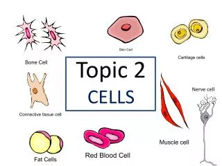
Review Topic 2. Cell membrane defines cell. Cell membrane separates living cell from aqueous environment thin barrier = 8nm thick Controls traffic in & out of the cell allows some substances to cross more easily than others hydrophobic (nonpolar) vs. hydrophilic (polar).

E N D
Cell membrane defines cell Cell membrane separates living cell from aqueous environment thin barrier = 8nm thick Controls traffic in & out of the cell allows some substances to cross more easily than others hydrophobic (nonpolar) vs. hydrophilic (polar)
Many Functions of Membrane Proteins “Channel” Outside Plasma membrane Inside Transporter Enzymeactivity Cell surfacereceptor “Antigen” Cell adhesion Cell surface identity marker Attachment to thecytoskeleton
Membrane carbohydrates Play a key role in cell-cell recognition ability of a cell to distinguish one cell from another antigens important in organ & tissue development basis for rejection of foreign cells by immune system
Simple Diffusion Move from HIGH to LOW concentration “passive transport” no energy needed movement of water diffusion osmosis
Facilitated Diffusion Diffusion through protein channels channels move specific molecules across cell membrane no energy needed HIGH LOW facilitated = with help open channel = fast transport “The Bouncer”
Concentration of water Direction of osmosis is determined by comparing total solute concentrations Hypertonic - more solute, less water, low water potential Hypotonic - less solute, more water, high water potential Isotonic - equal solute, equal water hypotonic hypertonic water net movement of water
Managing water balance Cell survival depends on balancing water uptake & loss freshwater balanced saltwater
Aquaporins Water moves rapidly into & out of cells evidence that there were water channels protein channels allowing flow of water across cell membrane 1991 | 2003 Peter Agre John Hopkins Roderick MacKinnon Rockefeller
Do you understand Osmosis… .05 M .03 M Cell (compared to beaker) hypertonic or hypotonic Beaker (compared to cell) hypertonic or hypotonic Which way does the water flow? in or out of cell
Active Transport Cells may need to move molecules against concentration gradient conformational shape change transports solute from one side of membrane to other protein “pump” “costs” energy = ATP LOW HIGH conformationalchange ATP “The Doorman”
Endocytosis fuse with lysosome for digestion phagocytosis non-specificprocess pinocytosis triggered bymolecular signal receptor-mediated endocytosis
Enzymes vocabulary substrate reactant which binds to enzyme enzyme-substrate complex: temporary association product end result of reaction active site enzyme’s catalytic site; substrate fits into active site active site products substrate enzyme
Properties of enzymes Reaction specific each enzyme works with a specific substrate chemical fit between active site & substrate H bonds & ionic bonds Not consumed in reaction single enzyme molecule can catalyze thousands or more reactions per second enzymes unaffected by the reaction Affected by cellular conditions any condition that affects protein structure temperature, pH, salinity
Induced fit model More accurate model of enzyme action 3-D structure of enzyme fits substrate substrate binding cause enzyme to change shape leading to a tighter fit “conformational change” bring chemical groups in position to catalyze reaction
Protein denaturation Unfolding a protein conditions that disrupt H bonds, ionic bonds, disulfide bridges temperature pH salinity alter 2° & 3° structure alter 3-D shape destroys functionality some proteins can return to their functional shape after denaturation, many cannot
Enzyme concentration What’shappening here?! reaction rate enzyme concentration
Substrate concentration What’shappening here?! reaction rate substrate concentration
Temperature 37° What’shappening here?! reaction rate temperature
pH What’shappening here?! pepsin trypsin pepsin reaction rate trypsin 0 1 2 3 4 5 6 7 8 9 10 11 12 13 14 pH
Salinity What’shappening here?! reaction rate salt concentration
Compounds which help enzymes Activators cofactors non-protein, small inorganic compounds & ions Mg, K, Ca, Zn, Fe, Cu bound within enzyme molecule coenzymes non-protein, organic molecules bind temporarily or permanently toenzyme near active site many vitamins NAD (niacin; B3) FAD (riboflavin; B2) Coenzyme A Fe inhemoglobin Mg inchlorophyll
Compounds which regulate enzymes Inhibitors molecules that reduce enzyme activity competitive inhibition noncompetitive inhibition irreversible inhibition feedback inhibition
Allosteric regulation Conformational changes by regulatory molecules inhibitors keeps enzyme in inactive form activators keeps enzyme in active form Conformational changes Allosteric regulation
Cooperativity Substrate acts as an activator substrate causes conformational change in enzyme induced fit favors binding of substrate at 2nd site makes enzyme more active & effective hemoglobin • Hemoglobin • 4 polypeptide chains • can bind 4 O2; • 1st O2 binds • now easier for other 3 O2 to bind
Feedback inhibition Example synthesis of amino acid, isoleucine from amino acid, threonine isoleucine becomes the allosteric inhibitor of the first step in the pathway as product accumulates it collides with enzyme more often than substrate does threonine isoleucine
Interphase 90% of cell life cycle cell doing its “everyday job” produce RNA, synthesize proteins/enzymes prepares for duplication if triggered I’m working here! Time to divide& multiply!
Prophase Chromatin condenses visible chromosomes chromatids Centrioles move to opposite poles of cell animal cell Protein fibers cross cell to form mitotic spindle microtubules actin, myosin coordinates movement of chromosomes Nucleolus disappears Nuclear membrane breaks down green = key features
Transition to Metaphase Prometaphase spindle fibersattach to centromeres creating kinetochores microtubules attach at kinetochores connect centromeres to centrioles chromosomes begin moving green = key features
Metaphase Chromosomes align along middle of cell metaphase plate meta = middle spindle fibers coordinate movement helps to ensure chromosomes separate properly so each new nucleus receives only 1 copy of each chromosome green = key features
Anaphase Sister chromatids separate at kinetochores move to opposite poles pulled at centromeres pulled by motor proteins “walking”along microtubules actin, myosin increased production of ATP by mitochondria Poles move farther apart polar microtubules lengthen green = key features
Telophase Chromosomes arrive at opposite poles daughter nuclei form nucleoli form chromosomes disperse no longer visible under light microscope Spindle fibers disperse Cytokinesis begins cell division green = key features
Cytokinesis Animals constriction belt of actin microfilaments around equator of cell cleavage furrow forms splits cell in two like tightening a draw string
Cytokinesis in Plants Plants cell plate forms vesicles line up at equator derived from Golgi vesicles fuse to form 2 cell membranes new cell wall laid down between membranes new cell wall fuses with existing cell wall