Download
1 / 42
610 likes | 1.96k Views
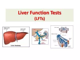
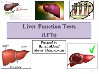
Abnormal LFTs. Michele Ritter Argy Resident – February, 2007. Liver Function Test. Albumin Bilirubin: Total Bilirubin Direct Bilirubin (conjugated bilirubin) Serum aminotransferases Aspartate aminotransferase (AST) Alanine aminotransferase (ALT) Alkaline Phosphatase Prothrombin time.

E N D
Abnormal LFTs Michele Ritter Argy Resident – February, 2007
Liver Function Test • Albumin • Bilirubin: • Total Bilirubin • Direct Bilirubin (conjugated bilirubin) • Serum aminotransferases • Aspartate aminotransferase (AST) • Alanine aminotransferase (ALT) • Alkaline Phosphatase • Prothrombin time
Albumin • Synthesized in the liver • Production is controlled by multiple factors including nutritional status, serum oncotic pressure, cytokines, and hormones • A serum albumin may be reflection of the synthetic function of the liver.
Bilirubin • Used to determine liver’s ability to clear endogenous/exogenous substances from the circulation • Indirect (unconjugated) bilirubin • Elevated with hemolysis, hepatic disease • Direct (conjugated) bilirubin • Elevated with biliary obstruction and hepatocellular disease. • Jaundice usually develops with a bilirubin ≥ 3 mg/dL
Aminotransferases • Hepatic enzymes that are usually intracellular, but are released from hepatocytes with hepatocellular injury. • Includes aspartate aminotransferase (AST) and alanine aminotransferase (ALT) • AST/ALT ratio • Normal is 0.8 • In alcoholic hepatitis, is usually > 2
Alkaline Phosphatase • A group of enzymes that catalyze the hydrolysis of a large number of organic phosphate esters. • In liver, believed to play an active role in down-regulating the secretory activities of the intrahepatic biliary epithelium • Found in: • Liver • Bone • intestine • First trimester placenta • Kidney • Gamma-glutamyl transpeptidase (GGT): • Liver origin: Elevated GGT • Bone origin: Normal GGT
Prothrombin Time (PT) • Liver is in charge of the synthesis of many clotting factors : • Factor I (fibrinogen) • Factor II (prothrombin) • Factor V • Factor VII • Factor IX • Factor X • Factors XII and XIII • Elevated PT may be reflection of decreased synthetic activity of liver.
Assessing the patient with abnormal Liver Function Tests • Most of the time, the cause of elevated LFTs can be illicited without invasive testing (biopsy) • If no cause of abnormality is found, most frequently the cause is alcohol liver disease, steatosis, or steatohepatitis • Certain patterns exist with LFTs • Hepatocellular Injury: Very high AST, ALT with mild/moderately elevated alkaline phosphatase. • Cholestatis: mild/moderately elevated AST/ALT with very high alkaline phosphatase • Bilirubin can be elevated with both combinations.
Hepatocellular Injury • Medications: • History: Need to assess temporal relationship with drug, see if patient improves once medication removed • NSAIDs, antibiotics, statins, anti-tuberculosis medications, anti-epileptic drugs, acetaminophen • Frequently cause isolated elevated aminotransferases • Acetaminophen overdose • Toxicity is likely to occur with single ingestions greater than 250 mg/kg or those greater than 12 g over a 24-hour period • AST/ALT elevations is first sign of liver damage (usually 24-hours after ingestion) • Alcohol Use: • Frequently have AST:ALT ratio ≥ 2:1 • History: Need accurate assessment of alcohol intake, including CAGE questions.
Hepatocellular Injury • Hepatitis A: • Acute infection • History: travel, recent outbreak, MSM; nausea, vomiting, jaundice • Labs: Hepatitis A IgM, frequent elevated bilirubin • Hepatitis B: • Can be acute or chronic • History: See if patient from Asia, Subsaharan Africa; Sexual history, Drug use • Labs: Hepatitis B surface antigen, surface antibody, core antibody • Hepatitis C: • History: IV drug abuse, blood transfusion prior to 1992, Sexual history, Tattoos • Labs: Hepatitis C antibody (Hepatitis C viral load if HIV positive or immunocompromised) • HIV: • Often causes isolated elevated aminotransferases • History: Sexual History, IV drug use • Labs: HIV Antibody test (ELISA with reflex Western Blot)
Hepatocellular Injury • Hereditary Hemochromatosis • History: Family history of liver disease? Diabetes? Heart Failure? Bronze skin? • Labs: • Serum iron, TIBC • Calculate iron saturation = serum iron/TIBC • If iron saturation > 45%, check ferritin • Ferritin • If > 400 ng/mL in men, or > 300 ng/mL in women, then need to check liver biopsy or genetic testing • Liver biopsy • Homozygous hereditary hemochromatosis if iron index > 1.9 • If under age 40, and positive genetic testing, no biopsy needed. • Genetic Testing
Hepatocellular Injury • Hepatic steatosis/Non-alcoholic steatohepatitis (NASH) • Increase in AST/ALT are usually less than 4-fold. • Ratio of AST/ALT is usually < 1 • History: Female, obesity, diabetes • Labs: • Labs to rule out other causes of hepatitis • Abdominal Ultrasound: look for fatty infiltration of liver
Hepatocellular Injury • Autoimmune Hepatitis • History: Young to middle-aged female • Labs: • Serum protein electrophoresis (SPEP) – if polyclonal increase in gamma globulin • Anti-nucleur antibody: Positive • Anti-smooth-muscle antibody (SMA) • Liver biopsy: should be performed if the above are negative, but autoimmune hepatitis still suspected.
Hepatocellular Injury • Alpha-1-antitrypsin deficiency • History: Family history, emphysema, young age • Labs: • Alpha-1-antitrypsin level/phenotype • Treatment: • Intravenous alpha-1 antiprotease helps with lung disease, but liver transplant is ultimately only treatment for liver disease.
Hepatocellular Injury • Wilson’s Disease • A genetic disorder of biliary copper excretion • History: Age (usually age 5 – 25, but up to age 40), family history of liver disease; neuropsychiatric disease • Evaluation: • Serum ceruloplasmin: Low • Opthalmologist: Exam for Kayser-Fleisher rings • 24-hour urine copper • Liver biopsy: Evaluate liver copper levels • Treatment: • Copper chelating agents • Zinc • In some cases, ultimately liver transplant
Hepatocellular Injury • Shock Liver (ischemic hepatitis) • Etiology: Shock, severe hypotension • Severely elevated AST/ALT (50 times normal) • Treatment: Re-establish good blood pressure/perfusion. • Prognosis: Usually patients recover, but can progress to fulminant liver failure requiring transplant.
Hepatocellular Injury • Non-Hepatic Causes • Usually only mild increase in AST/ALT • Muscle disorders • Hypothyroidism/Hyperthyroidism • Celiac Disease • Adrenal Insufficiency • Anorexia nervosa
Hepatocellular Injury • What if work-up is negative and AST/ALT remain elevated? • Observe: • Patients with two-fold or less increase in AST/ALT and no hyperbilirubinemia • Liver Biopsy • Patients with > two-fold increase in AST/ALT, or abnormalities of other liver function tests.
Cholestatic Pattern • Predominantly elevated alkaline phosphatase • Need to check GGT to see if bone or liver in origin • Blood types O and B: can have elevated serum alkaline phosphatase after eating a fatty meal due to an influx of intestinal alkaline phosphatase • Need to determine if the cholestasis is intrahepatic or extrahepatic in origin.
Cholestatic Pattern - Intrahepatic • Drugs: • Anabolic steroids, contraceptives, antibiotics • Total parenteral nutrition (TPN) • Cirrhosis: • Viral hepatitis (Hepatitis B, C) • Alcohol hepatitis
Cholestatic Pattern - Intrahepatic • Primary Biliary Cirrhosis • Autoimmune disease • Predominately in women, usually ages 35-65 • May have history of other autoimmune disease • Symptoms: Prurutis, fatigue, hyperpigmentation, musculoskeletal complaints • Labs: • RUQ Ultrasound • Anti-mitochondrial antibody • Liver biopsy to verify diagnosis
Cholestatic Pattern – Both Intrahepatic and Extrahepatic • Primary Sclerosing Cholangitis • chronic progressive disorder of unknown etiology that is characterized by inflammation, fibrosis, and stricturing of medium size and large ducts in the intrahepatic and extrahepatic biliary tree • ~ 90% have inflammatory bowel disease, especially ulcerative colitis • Symptoms: Pruritus, fatigue, RUQ pain • Diagnosis: • Ultrasound • Cholangiogram: multifocal stricturing and dilation of intrahepatic and/or extrahepatic bile ducts • Prognosis: • Poor; average life expectancy after diagnosis is ~12 years • 10-15% risk of developing cholangiocarcinoma • Liver transplant is ultimate only treatment
Cholestatic Pattern - Extrahepatic • Choledocholithiasis(gall stones!) • History: • The 3 F’s • RUQ colicky abdominal pain • Diagnosis/Treatment: Ultrasound, ERCP (to remove stones) • Malignancy • Cholangiocarcinoma • Pancreatic • Metastatic cancer • Diagnosis: Ultrasound, MRCP • Treatment: ERCP, biliary stent
Cholestatic Pattern - Extrahepatic • Chronic Pancreatitis • History: Recurrent pancreatitis • Symptoms: Abdominal pain, frequently referred to back • HIV Cholangiopathy • Usually seen in AIDS patients with CD4 count well below 100/mm3 • Usually caused by: Cryptosporidium. Microsporidium, CMV • Symptoms: RUQ pain, Diarrhea, Occassional fever, Occassional jaundice • Diagnosis: • ERCP • Cholangiography – shows multifocal strictures of extrahepatic biliary tree
Isolated Hyperbilirubinemia • Unconjugated (indirect) hyperbilirubinemia • Overproduction of bilirubin • Hemolysis • Dubin-Johnson Syndrome and Rotor Syndrome • Decrease in uptake, conjugation, or excretion of bilirubin • Increased unconjugated (indirect) bilirubin • Liver Disease
Isolated Unconjugated Hyperbilirubinemia • Drugs • Probenecid, Rifampicin • Gilbert’s Disease • Autosomal recessive disorder • 3 to 7 % of population • Most common in white males • Jaundice, increased unconjugated bilirubin (always < 6) • Occurs when patient under stress/infection • Crigler-Najjar type II • Caused by gene mutation • Reduced activity of Bilirubin UDP glucuronosyl
Hepatocellular Injury – mostly AST/ALT Drugs Alcohol hepatitis Hepatitis A Hepatitis B Hepatitis C Steatohepatitis (NASH) Autoimmune hepatitis Wilson’s Disease Hereditary Hemochromatosis Alpha-1 antitrypsin deficiency SUMMARY
EXTRAHEPATIC Gall stones Primary Sclerosing Cholangitis Malignancy Chronic pancreatitis HIV cholangiopathy Cholestatic Pattern INTRAHEPATIC Drugs Hepatitis A, B, C Alcoholic hepatitis TPN Primary Sclerosing Cholangitis Primary Biliary Cirrhosis SUMMARY
SUMMARY • Isolated elevated indirect (unconjugated) bilirubin • Hemolysis • Drugs • Gilbert’s Disease • Crigler-Najjar type II
Scenario # 1 • A 43-year old woman who has consumed a pint of 80-proof whiskey daily for 18 years presents with right upper quadrant pain. The pain began approximately a week ago and has been transiently relieved by her taking two extra-strength acetaminophen tablets every 4 hours for the past 4 days. She has had some nausea and vomiting but no fever. There is no history of jaundice or cholelithiasis. The patient used intravenous drugs and shared needles during her late teen years.
Scenario # 1 • Physical Exam • Enlarged tender liver that percusses to 17 cm in the right midclavicular line and a tattoo on the right buttock • Labs: • Bilirubin: 2 mg/dL AST: 3800 Alk. Phos: 198 PT: normal
Scenario #1 • The most likely diagnosis is: (A) Alcoholic hepatitis (B) Acute cholecystitis (C) Acetaminophen hepatotoxicity (D) Acute viral hepatitis B (E) Acute viral hepatitis C
Scenario # 2 • A 54-year old asymptomatic man volunteers to donate blood and is found to have elevated aminotransferase levels. He has no known medical problems and no history of hepatitis. He drinks no alcohol, takes no medications, and has not seen a physician in more than 10 years. He is active, works as a truck driver, and has noted no change in his physical condition. He has no family history of liver disease.
Scenario # 2 • Physical Exam: • Obesity – Ht: 5’ 10”, 115 kg • Labs: • AST: 45 ALT: 85 Alk. Phos: 90 • Hepatitis serologies (A, B, C): negative • ESR: normal ANA: negative • Smooth muscle antibody: negatie • Total chol: 260 LDL; 225 Triglycerides: 830 • Liver biopsy: • Large-droplet steatosis without significant inflammatory reaction and no fibrosis. Ultrasonography shows a mildly enlarged fatty liver.
Scenario # 2 • The appropriate management of this patient would be: • Interferon therapy for presumed chronic non-B, non-C hepatitis • Alcohol rehabilitation and counseling • Weight loss and therapy for hyperlipidemia • Endoscopic retrograde cholangiopancreatography (ERCP) to evaluate the biliary tree • Corticosteroid therapy
Scenario # 3 • A 43-year old woman complains of itching that keeps her awake at night. Physical examination is normal except for the liver, which is felt 7 cm below the right costal margin. • CBC is normal • Creatinine: 0.8 mg/dL, Bilirubin: 0.6 mg/dL • ALT: 78 U/L, Albumin: 4.2 g/dL • Alkaline Phosphatase: 450 U/L • Cholangiogram: normal
Scenario # 3 • Which test would be most accurate in diagnosing her underlying disorder? (A) Serum protein electrophoresis (B) Anti-Smooth Muscle Antibody (C) Antimitochondrial antibody (D) Technetium-99m liver-spleen scan (E) Endoscopic retrograde cholangiopancreatography (ERCP)