Download
1 / 39
790 likes | 1.81k Views
NEONATAL CHOLESTASIS. Gregory J. Semancik, M.D. Major, Medical Corps, U.S. Army Fellow, Pediatric Gastroenterology and Nutrition Walter Reed Army Medical Center. OBJECTIVES. Know the differential diagnosis for neonatal cholestasis.

E N D
NEONATAL CHOLESTASIS • Gregory J. Semancik, M.D. • Major, Medical Corps, U.S. Army • Fellow, Pediatric Gastroenterology and Nutrition • Walter Reed Army Medical Center
OBJECTIVES • Know the differential diagnosis for neonatal cholestasis. • Understand how to evaluate the neonate with conjugated hyperbilirubinemia. • Know the therapeutic management of neonates with cholestasis.
DEFINITION • Neonatal cholestasis is defined as conjugated hyperbilirubinemia developing within the first 90 days of extrauterine life. • Conjugated bilirubin exceeds 1.5 to 2.0 mg/dl. • Conjugated bilirubin generally exceeds 20% of the total bilirubin.
ETIOLOGIES • Basic distinction is between: • Extrahepatic etiologies • Intrahepatic etiologies
EXTRAHEPATIC ETIOLOGIES • Extrahepatic biliary atresia • Choledochal cyst • Bile duct stenosis • Spontaneous perforation of the bile duct • Cholelithiasis • Inspissated bile/mucus plug • Extrinsic compression of the bile duct
INTRAHEPATIC ETIOLOGIES • Idiopathic • Toxic • Genetic/Chromosomal • Infectious • Metabolic • Miscellaneous
INTRAHEPATIC ETIOLOGIES • Idiopathic Neonatal Hepatitis • Toxic • TPN-associated cholestasis • Drug-induced cholestasis • Genetic/Chromosomal • Trisomy 18 • Trisomy 21
INTRAHEPATIC ETIOLOGIES • Infectious • Bacterial sepsis (E. coli, Listeriosis, Staph. aureus) • TORCHES • Hepatitis B and C • Varicella • Coxsackie virus • Echo virus • Tuberculosis
INTRAHEPATIC ETIOLOGIES • Metabolic • Disorders of Carbohydrate Metabolism • Galactosemia • Fructosemia • Glycogen Storage Disease Type IV • Disorders of Amino Acid Metabolism • Tyrosinemia • Hypermethioninemia
INTRAHEPATIC ETIOLOGIES • Metabolic (cont.) • Disorders of Lipid Metabolism • Niemann-Pick disease • Wolman disease • Gaucher disease • Cholesterol ester storage disease • Disorders of Bile Acid Metabolism • 3B-hydroxysteroid dehydrogenase/isomerase • Trihydroxycoprostanic acidemia
INTRAHEPATIC ETIOLOGIES • Metabolic (cont.) • Peroxisomal Disorders • Zellweger syndrome • Adrenoleukodystrophy • Endocrine Disorders • Hypothyroidism • Idiopathic hypopituitarism
INTRAHEPATIC ETIOLOGIES • Metabolic (cont.) • Miscellaneous Metabolic Disorders • Alpha-1-antitrypsin deficiency • Cystic fibrosis • Neonatal iron storage disease • North American Indian cholestasis
INTRAHEPATIC ETIOLOGIES • Miscellaneous • Arteriohepatic dysplasia (Alagille syndrome) • Nonsyndromic paucity of intrahepatic bile ducts • Caroli’s disease • Byler’s disease • Congenital hepatic fibrosis
INTRAHEPATIC ETIOLOGIES • Miscellaneous (cont.) • Familial benign recurrent intrahepatic cholestasis • Hereditary cholestasis with lymphedema (Aagenaes) • Histiocytosis X • Shock • Neonatal lupus
COMMON ETIOLOGIES • Premature infants • Sepsis/Acidosis • TPN-associated • Drug-induced • Idiopathic neonatal hepatitis • Extrahepatic biliary atresia • Alpha-1-antitrypsin deficiency • Intrahepatic cholestasis syndromes
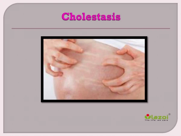
CLINICAL PRESENTATION • Jaundice • Scleral icterus • Hepatomegaly • Acholic stools • Dark urine • Other signs and symptoms depend on specific disease process
GOALS OF TIMELY EVALUATION • Diagnose and treat known medical and/or life-threatening conditions. • Identify disorders amenable to surgical therapy within an appropriate time-frame. • Avoid surgical intervention in intrahepatic diseases.
EVALUATION • Basic evaluation • History and physical examination (includes exam of stool color) • CBC and reticulocyte count • Electrolytes, BUN, creatinine, calcium, phosphate • SGOT, SGPT, GGT, alkaline phosphatase • Total and direct bilirubin • Total protein, albumin, cholesterol, PT/PTT
EVALUATION • Tests for infectious causes • Indicated cultures of blood, urine, CSF • TORCH titers, RPR/VDRL • Urine for CMV • Hepatitis B and C serology • Ophthalmologic examination
EVALUATION • Metabolic work-up • Protein electrophoresis, alpha-1-antitrypsin level and phenotype • Thyroid function tests • Sweat chloride • Urine/serum amino acids • Review results of newborn metabolic screen • Urine reducing substances • Urine bile acids
EVALUATION • Radiological evaluation • Ultrasonography • Patient should be NPO to increase likelihood of visualizing the gallbladder • Feeding with exam may demonstrate a functioning gallbladder • Hepatobiliary scintigraphy • Premedicate with phenobarbital 5mg/kg/d for 3-5 days
EVALUATION • Invasive studies • Duodenal intubation • Percutaneous liver biopsy • Percutaneous transhepatic cholangiography • Endoscopic retrograde cholangiopancreatography (ERCP) • Exploratory laparotomy with intraoperative cholangiogram
EXTRAHEPATIC BILIARY ATRESIA • Generally acholic stools with onset at about 2 weeks-old • Average birth weight • Hepatomegaly with firm to hard consistency • Female predominance • No well-documented familial cases
EXTRAHEPATIC BILIARY ATRESIA • Increased incidence of polysplenia syndrome and intra-abdominal vascular anomalies • Normal uptake on radionucleotide scan with absent excretion • Biopsy shows bile duct proliferation, bile plugs, portal or perilobular fibrosis and edema, and intact lobular structure
IDIOPATHIC NEONATAL HEPATITIS • Generally normal stools or acholic stools with onset at one month-old • Low birth weight • Normal liver on exam or hepatomegaly with normal to firm consistency • Male predominance • Familial cases (15-20%)
IDIOPATHIC NEONATAL HEPATITIS • Impaired uptake on radionucleotide scan with normal excretion • Biopsy shows intralobular inflammation with focal hepatocellular necrosis and disruption of the hepatic architecture. No alteration of the bile ducts. Giant cell transformation occurs but is non-specific.
ALPHA-1-ANTITRYPSIN DEFICIENCY • Alpha-1-antitrypsin makes up 90% of alpha-1-globulin fraction • Associated with PiZZ (about 10-20% will have liver disease) and rarely with PiSZ and PiZ-null phenotypes • Biopsy shows hepatocellular edema, giant cell transformation, necrosis, and pseudoacinar transformation.
ALPHA-1-ANTITRYPSIN DEFICIENCY • Biopsy also shows accumulation of PAS-positive, diastase-resistant globules in the cytoplasm of periportal hepatocytes. • Varying degrees of fibrosis correlate with disease prognosis.
INTRAHEPATIC CHOLESTASIS SYNDROMES • Includes several diagnostic entities. • Biopsies show cholestasis. May show paucity of intrahepatic bile ducts, giant cell transformation, and/or fibrosis.
TREATMENT • Surgical • Kasai procedure for biliary atresia • Limited bile duct resection and re-anastomosis • Choledochal cyst excision • Cholecystectomy • Liver transplantation
KASAI PROCEDURE • Performed for biliary atresia that is not surgically correctable with excision of a distal atretic segment. • Roux-en-Y portoenterostomy • Bile flow re-established in 80-90% if performed prior to 8 weeks-old. • Bile flow re-established in less than 20% if performed after 12 weeks-old
KASAI PROCEDURE • Success of the operation is dependent on the presence and size of ductal remnants, the extent of the intrahepatic disease, and the experience of the surgeon. • Complications are ascending cholangitis and reobstruction as well as failure to re-establish bile flow.
LIVER TRANSPLANTATION • Survival rates approach 80% at 1 year and 70% at 5 years. • Biliary atresia is the most common indication for transplant and may be the initial treatment when detected late or may be used as a salvage procedure for a failed Kasai. • Used early in cases of tyrosinemia.
TREATMENT • Medical management • Nutritional support • Treatment of pruritus • Choleretics and bile acid-binders • Management of portal hypertension and its consequences
TREATMENT • Nutritional support • Adequate calories and protein • Supplement calories with medium chain triglycerides • Maintain levels of essential long-chain fatty acids • Treatment and/or prophylaxis for fat-soluble vitamin deficiencies (vitamins A, D, E, and K)
TREATMENT • Nutritional support (cont.) • Supplemental calcium and phosphate when bone disease is present • Prophylaxis for zinc deficiency • Low-copper diet as poorly excreted • Sodium restriction when ascites present
TREATMENT • Treatment of pruritus • Bile acid-binders: cholestyramine, cholestipol • Ursodeoxycholic acid • Phenobarbital as a choleretic • Naloxone • Rifampin
TREATMENT • Management of portal hypertension and its consequences • Variceal bleeding • Fluid rescuscitation • Blood products • Sclerotherapy • Balloon tamponade • Portovenous shunting • Propanolol
TREATMENT • Management of portal hypertension and its consequences (cont.) • Ascites • Sodium restriction • Diuretics: spironolactone, furosemide • Albumin • Paracentesis • Thrombocytopoenia managed with platelet infusions when clinically indicated