Download
1 / 19
210 likes | 892 Views
Scanning Probe Microscopy. Colin Folta Matt Hense ME381R 11/30/04. Outline . Background and History AFM MFM EFM SThM STM SHFM SNOM. Background . First scanning probe microscope invented in 1981 by Binning and Roher Wide range of applications Topography/Atomic Structure

E N D
Scanning Probe Microscopy Colin Folta Matt Hense ME381R 11/30/04
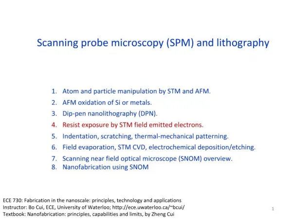
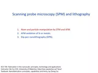
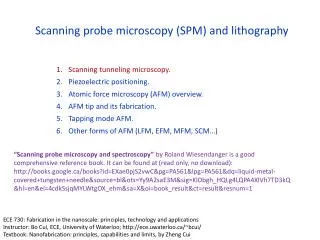
Outline • Background and History • AFM • MFM • EFM • SThM • STM • SHFM • SNOM
Background • First scanning probe microscope invented in 1981 by Binning and Roher • Wide range of applications • Topography/Atomic Structure • Magnetic/Electric fields • Surface temperatures
Branches of Scanning Probe Microscopy http://spm.phy.bris.ac.uk/
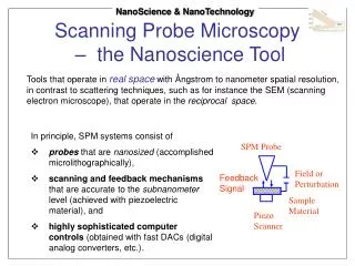
Operation • Scanning probe microscopes operate by detecting the deflection in the cantilever • Modern scanning probe microscopes use a split photo diode to detect the deflection http://spm.phy.bris.ac.uk/
Atomic Force Microscopy (AFM) • Most widely used branch of scanning probe microscopy • Operates by measuring the interaction force between the tip and sample
AFM Operation Modes • Contact Mode • Tip remains in the repulsive regime of the inter-molecular force curve • Tapping Mode • Tip is oscillated at a high frequency • Deflections in the oscillations are observed • Non-Contact Mode • Tip is oscillated outside of the repulsive regime
Image Defects • Broadening • Occurs when feature is roughly the same size as the radius of curvature • Side wall of tip comes into contact before the tip itself • Compression • The forces involved actually change the shape of the specimen (ex. DNA)
Image Defects Cont. • Aspect Ratio • Steep walled features become distorted • The tip can not follow a perfectly vertical wall http://spm.phy.bris.ac.uk/
Magnetic Force Microscopy (MFM) • Coated with a magnetic covering • Two modes of operation • Non-vibrating for larger magnetic fields • Vibrating for weaker fields that require a greater sensitivity
MFM Cont. • Uses a two pass technique • First pass finds topography of sample • Second pass finds the magnetic field • On the second pass tip is kept at a constant height http://www.ntmdt.ru/SPM-Techniques/SPM-Methodology/ Magnetic_Force_Microscopy_MFM/text45.html
Electrostatic Force Microscopy (EFM) • A bias is used to create an electrostatic field between the tip of the probe and the sample • Two uses • Determine which regions are conducting and which are insulating • Determine the electric potential at different points
Scanning Tunneling Microscopy (STM) • Electrons are transferred between the tip and the sample due to overlapping orbitals • A net transfer can be sustained by applying a voltage across the gap • Change in current is a result of a change in the tip-sample separation http://stm1.phys.cmu.edu/stm/si5x5s.gif http://www.d.umn.edu/~jmaps/stm1.html
STM Modes of Operation • Constant Current • Maintain a constant tunneling current by adjusting the separation • Constant Height • Maintain a constant height and measure the current change
Scanning Thermal Microscopy (SThM) • Thermocouple is placed on the tip of the probe • Combined with AFM, SThM can associate thermal properties with surface features • By heating the tip ~30K higher than the sample, local thermal conductivity can be determined • Thermocouple can be used conventionally to measure temperature distribution along the sample
Scanning Near Field Optical Microscopy (SNOM) • Typical optical microscopes • Limited by the Abbe diffraction barrier • Resolution equal to one half of the wavelength of the light http://molebio.iastate.edu/~p_haydon/nsom.html
SNOM Cont. • SNOM • Uses a very small aperture • Keeps the specimen in the near field regime • Resolution is determined by the aperture diameter http://spm.phy.bris.ac.uk/
Shear Force Microscopy (ShFM) • Probe oscillates parallel to the specimen • Oscillation changes because of Van der Waals interactions. Topography can be determined from these changes. • Advantages • More rigid set up • “Jump to contact” problem is almost eliminated • Disadvantages • Can be very difficult to set up • Probe tip is very hard to reproduce reliably