Download
1 / 1
10 likes | 97 Views
A Quantitative Study of Bioenergetics in Skeletal Muscle Lacking Carbonic Anhydrase III Using 31P Magnetic Resonance Spectroscopy. PI: Krista Vandenborne; University of Florida NIH Grant AR45394.

E N D
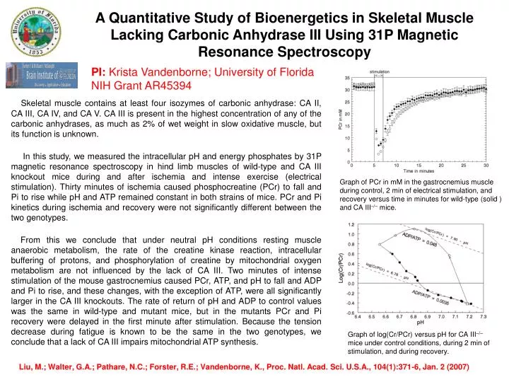
A Quantitative Study of Bioenergetics in Skeletal Muscle Lacking Carbonic Anhydrase III Using 31P Magnetic Resonance Spectroscopy PI: Krista Vandenborne; University of Florida NIH Grant AR45394 Skeletal muscle contains at least four isozymes of carbonic anhydrase: CA II, CA III, CA IV, and CA V. CA III is present in the highest concentration of any of the carbonic anhydrases, as much as 2% of wet weight in slow oxidative muscle, but its function is unknown. In this study, we measured the intracellular pH and energy phosphates by 31P magnetic resonance spectroscopy in hind limb muscles of wild-type and CA III knockout mice during and after ischemia and intense exercise (electrical stimulation). Thirty minutes of ischemia caused phosphocreatine (PCr) to fall and Pi to rise while pH and ATP remained constant in both strains of mice. PCr and Pi kinetics during ischemia and recovery were not significantly different between the two genotypes. From this we conclude that under neutral pH conditions resting muscle anaerobic metabolism, the rate of the creatine kinase reaction, intracellular buffering of protons, and phosphorylation of creatine by mitochondrial oxygen metabolism are not influenced by the lack of CA III. Two minutes of intense stimulation of the mouse gastrocnemius caused PCr, ATP, and pH to fall and ADP and Pi to rise, and these changes, with the exception of ATP, were all significantly larger in the CA III knockouts. The rate of return of pH and ADP to control values was the same in wild-type and mutant mice, but in the mutants PCr and Pi recovery were delayed in the first minute after stimulation. Because the tension decrease during fatigue is known to be the same in the two genotypes, we conclude that a lack of CA III impairs mitochondrial ATP synthesis. Graph of PCr in mM in the gastrocnemius muscle during control, 2 min of electrical stimulation, and recovery versus time in minutes for wild-type (solid ) and CA III–/– mice. Graph of log(Cr/PCr) versus pH for CA III–/– mice under control conditions, during 2 min of stimulation, and during recovery. Liu, M.; Walter, G.A.; Pathare, N.C.; Forster, R.E.; Vandenborne, K., Proc. Natl. Acad. Sci. U.S.A., 104(1):371-6, Jan. 2 (2007)