Download
1 / 9
90 likes | 178 Views
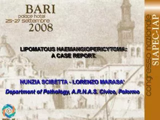
IUXTA-ARTICULAR MYXOMA : A CASE REPORT. NUNZIA SCIBETTA - LORENZO MARASA’ Department of Pathology, A.R.N.A.S. Civico, Palermo. INTRODUCTION.

E N D
IUXTA-ARTICULAR MYXOMA : A CASE REPORT NUNZIA SCIBETTA - LORENZO MARASA’ Department of Pathology, A.R.N.A.S. Civico, Palermo
INTRODUCTION • Iuxta-articular myxoma (JAM), is a rare, benign soft tissue tumor that usually arises in the vicinity of a large joint ( knee, shoulder, elbow, ankle), and has histological features resembling a cellular myxoma. • The patients ranged in age from 16 to 83 years and present with a swelling or a mass that can be painful or tender. • Radiographic studies show a soft tissue mass that has similar imaging characteristics as intramuscular myxoma. • We report a case of JAM situated in the vicinity of a left temporo-mandibolare joint, because no cases has been reported before, in this seat, in the pathology literature.
METHODS • A 68 year old man underwent conservative excision of a enlarging, painful mass, arising in the vicinity of a left temporo-mandibular joint. • No history of trauma or surgery was reported. • The surgical specimen was fixed in buffered formalin, embedded in paraffin and stained with H&E. Histo-chemical (colloidal iron, alcian-blue and mucicarmine stains for stromal mucins (glycosaminoglycans), and immunohistochemical studies were performed.
RESULTS • Grossly the lesion of 3 cm of maximum diameter, was myxoid, soft, yellow-tan, and involved the subcuta-neous adipose tissue. • Histologically the lesion showed loosely arranged spin-dled fibroblast-like cells suspended in an abundant hy-povascular, rich myxoid matrix. • No cystic, ganglion-like spaces were seen. • Mitotic figures and necrosis are absent • The periphery of the tumor showed ill defined bounda-ries. • Colloidal iron, alcian blue and mucicarmine stains for stromal mucins confirmed the abundance of glycosami-noglycans in the myxoid matrix, and the spindled cells showed staining for CD34, vimentin, and no staining for S100 protein, desmin and actin.
2 1 2 Bland appearing spindle cells em-bedded in a hypovascular myxoid stroma JAM composed of fibroblast-like cells with hyperchromatic nuclei and delicate cytoplasmic processes
2 1 2 Cells with small cytoplasmic vacuo-les containing mucinous material, mild nuclear pleomorphism and hy-perchromasia Multiple small cystic spaces and clefts are seen in the myxoid stroma
1 2 2 1 . Immunohistochemically the cells stain for CD34 2 . There is no staining for desmin 3 . There is no staining for S100 pro-tein 3
CONCLUSIONS • The our lesion involved the subcutaneous adipose tissue in a man and presented as an enlarging, painful mass in the region of temporo-mandibular joint. • Microscopically bears a close resemblance to intramu-scular myxoma , but there are certain clinical and mi-croscopic differences between this lesion and JAM, because intramuscular myxoma tend to affect woman and occur primarly in the large muscles of the body, and tipically do not recur even when incompletely exci-sed.
CONCLUSIONS • JAM is likely a proliferative fibroblastic process with excess extracellular mucin formation as seen in intramuscular myxoma, early forms of ganglion cysts, and also in nodular fasciitis, another presumably reactive proliferation of fibroblasts. • JAM is a benign condition despite its frequent large size, its infiltrative growth pattern. Therefore complete conservative excision is the optimal treatment. MAIN REFERENCE Meis J.M., Enzinger F. : Juxta-articular myxoma. A Clinical and Pathologic Study of 65 cases. Human Pathol 23; 639-646; 1992