Slide Note
0 likes | 6 Views
MEN1 and MEN2 are rare autosomal dominant familial cancer syndromes with varied presentations and associated tumors. This study explores the genetic basis and prevalence of MEN, covering key manifestations, diagnostic techniques, and management strategies, including genetic testing and specialist multidisciplinary team involvement.

E N D
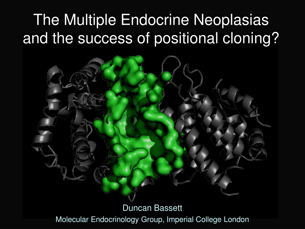
The Multiple Endocrine Neoplasias and the success of positional cloning? Duncan Bassett Molecular Endocrinology Group, Imperial College London
MEN1 and MEN2 Rare autosomal dominant familial cancer syndromes Prevalence 1 per 100,000 Presentation 5 to 81 years Prevalence 1 per 500,000 High penetrance 6/12 to 40 years MEN1 Parathyroid tumours Pancreatic islet cell tumours (NETs) Anterior pituitary tumours Adrenal cortical tumours Gut, thymic, bronchial carcinoids (NETs) Angiofibromas, lipoma collaginomas, meningiomas MEN 2A (60%) Medullary thyroid carcinoma Phaeochromocytoma Parathyroid hyperplasia MEN 2B (5%) Medullary thyroid carcinoma Phaeochromocytoma Marfanoid habitus Mucosal neuromas Ganglioneuromatosis/megacolon Isolated familial syndromes Hyperparathyroidism Prolactinomas/Acromegaly Carcinoids Isolated familial syndromes (35%) Familial MTC Familial Phaeo
Complex Multiple Endocrine Neoplasias MEN4 (CDKN1B) Parathyroid tumours Pituitary tumours McCune Albright (GNAS1) Thyroid nodular hyperplasia (TTX) Adrenal hyperplasia (Cushing's) Somatotrophinomas (Acromegaly) Hyperprolactinaemia Neurofibromatosis I (Neurofibromin) Phaeochromocytoma Hyperparathyroidism Carcinoids (NETs) (Medullary thyroid carcinoma) Von Hippel-Lindau (VHL) Phaeochromocytoma Pancreatic Islet cell tumour Thyroid tumours Parathyroid tumours Adrenal tumours Pituitary tumours Carney’s Complex (PRKAR1A) Familial isolated pituitary adenomas (FIPA) (AIP) Somatotrophinomas, prolactinomas Somatolactotrophinomas, Non-functional
Multiple Endocrine Neoplasia Type 1 Parathyroid (94%) Pancreas (40%) 43% 26% 2% 11% 14% 1% 3% Pituitary (29%) Cutaneous tumours Angiofibromas 88% Collaginomas 72% Lipomata 30% Associated tumours Carcinoid 4% Adrenocortical 5% Phaeochromocytoma 0.5% (Jensen RT 2008 Cancer 113:1807-1843; Verges B 2002 JCEM 87:457-465)
10Hyperparathyroidism 10Hyperparathyroidism (95%) Frequently the first presenting feature Differs from sporadic disease Early age of presentation peak 20-25 years Multiple gland hyperplasia rather than adenoma High recurrence rate (50% by 10 years) Presentation Hypercalcaemia Polyuria, polydipsia, nephrocalcinosis, renal stones Abdominal pain, N/V, constipation Dyspepsia, peptic ulceration, pancreatitis Osteofibrosacystica Psychiatric disturbance
Pancreatic endocrine tumours Single/multiple, benign/malignant, functional/non-functional Angiography CT scan Pancreatic neuroendocrine tumours NETs (50-100%) Non-functional (PPomas) asymptomatic can metastasize if >4cm Gastrinoma (ZE) Peptic ulceration, diarrhoea and steatorrhoea Co-secreting Mixed symptoms Insulinoma Hypoglycaemic symptoms, Hunger, Wt gain Glucagonoma NME, DM, Wt loss, diarrhoea, DVT/PE VIPoma Severe watery diarrhoea Somatostatinomas (80-100%) (48%) (22%) (14%) (5%) (2%) (Rare) (Jensen RT 2008 Cancer 113:1807-1843)
Anterior pituitary tumours Cushing’s Acromegaly Pituitary Adenomas (40%) Mean age of onset 40 years (5-83) Initial lesion in 17% and more frequent in females More aggressive than sporadic disease 85% macroadenomas and 37% invasive
Clinical features of pituitary adenomas Lactotrophinomas (Prolactinomas) (Prolactin producing 63% ) Menstrual irregularity, galactorrhoea, reduced libido, impotence, infertility Somatotrophinomas (Acromegaly) (GH producing 23%) Headache, sweating, ↑soft tissue, ↑hands and feet, prognathism, nerve compression, cutaneous fibromas, acanthosis nigricans, HT, LVH, cardiomyopathy, arrhythmias, colonic malignancy ACTHomas (Cushings disease) (ACTH producing 4%) Hirsuitism, centripedal obesity, buffalo hump, purple striae, HT, glucose intolerance, proximal myopathy, infertility, psychiatric problems Non Functional tumours (15%) Headache, bitemporal hemianopia, hypopituitarism, cranial nerve palsies menstrual irregularity, galactorrhoea, reduced libido, impotence, infertility Co-secreatory (9%)
Other MEN1 associated tumours Adrenocortical (20-50%) Clinically silent adrenal adenomas or hyperplasia <10% functional (Conns/Cushing’s) Rarely carcinomas Carcinoid Syndrome (NETs) (15%) 70% foregut (gastric 70%), thymic (15%), and bronchial (15%) Flushing, palpitations, wheezing, diarrhoea, tricuspid insufficiency and pulmonary stenosis
Dermatological features of MEN1 Benign lesions no treatment required Lipomas (3-34%) Multiple facial angiofibromas (5- 88%) Hypopigmented collagenomas (0-72%)
2012 Clinical practice guidelines Management by specialist MDT Endocrinologist, gastroenterologist, oncologist, endocrine surgeon, specialist histopathologist, interventional radiologist and clinical geneticist Genetic testing Index case (2x MEN1 associated tumours) Screen 1stdegree relatives at earliest opportunity (before biochemical testing) Tumour 10HPT Pancreatic NET Annually Ca, PTH Glu, Gut hormonesSurgery/PPI/SS analogues MRI/Endo US PRL, IGF-1 MRI Combined PFT Dopamine agonist, SS analogues Transsphenoidal hypophsectomy Thymic/bronchial Chest MRI Gastroscopy Carcinoids Endo US SS-scintigraphy Adrenal MRI Biochemical evaluation if >1cm Surgery is functional (10%) (Surgery if NF>4cm) 3 yearly Management Parathyroidectomy (Cinacalcet) (Surgery if NF >1cm) Pituitary Surgical resection Radio/Chemotherapy (Thakker RV et al (2012) JCEM 97:2990-3011)
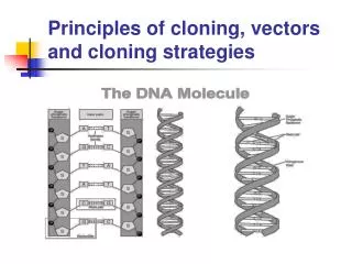
MEN1 is caused by loss of function mutations in a tumour suppressor gene that encodes MENIN
The MEN1 gene Chr 11 Located at chromosome 11q13 10 exons 2.8Kb mRNA transcript 610 amino acids protein Menin 6 alternative splice variants (5’ UTR) Menin is a tumour suppressor gene MEN1 is caused by Menin loss of function mutations 75% predict truncated or absent protein Rapid proteasome-degradation of mutant Menin Tumour specific loss of the normal MEN1 allele (LOH) “Knudsons two hit hypothesis” Over-expression of Menin in Ras-transformed cell lines reverts the transformed phenotype
Tumour suppressor genes and tumour formation Normal Individual MEN1 mutant gene carrier Susceptible cell with 2 functional MEN1 alleles Susceptible cell with 1 functional MEN1 allele Mutant MEN1 MEN1 MEN1 MEN1 Sporadic mutation with loss of 11q Susceptible cell with 1 functional MEN1 allele Susceptible cell no functional MEN1 alleles Mutant MEN1 MEN1 Tumours show LOH No tumour Tumour formation
Mutation are scattered throughout MEN1 gene 459 different germ line mutations identified 75% predict a truncated or absent protein No hot spots, scattered throughout coding region Nonsense Frameshift deletion/inertion Large deletions In frame deletion/insertion Missense 10% occur de novo 5-10% of patients do not have mutations in coding region 23% 41% 1% 6% 20% (Thakker RV 2010 Best Pract Clin Endo Metab 24:355; Human Gene Mutation Database Cardiff)
The MENIN protein 200 300 0 100 400 500 600 NLS1 NLSa NLS2 2 3 4 5 6 7 8 9 10 MENIN is a 67kDa protein, 3x NLS and binds DNA Nuclear localisation but cytoplasmic in dividing cells No homology or functional motifs Conserved from man to molluscs Drosophila 70% identity Expressed from earliest stages of embryogenesis Expressed ubiquitously in all adult tissues Expression inversely correlated with proliferation (cell cycle) Expression varies with the cell type Menin negatively regulates its own expression Somatostatin induces Menin expression Multiple protein interactions have been identified Precise cellular function of Menin remains uncertain The pattern of MENIN expression cannot explain the endocrine nature of associated tumours
MENIN interactions and function Two hybrid screening, co-immunoprecipitation and GST-pull-down studies (Chandrasekharappa SC 2003 J Int Med 253:606 ;Thakker RV 2010 Best Pract Clin Endo Metab 24:355)
Transcriptional regulation by Menin Interacts with JunD and C-Jun to suppresses transcription JunD/mSin3A/HDAC histone deacetylase recruitment Binds NF B (p50, p52 and p65) suppressing transcriptional activation Inhibits TGFb b and BMP-2 signally by binding Smad3 and Smad1/5 Menin is component of MLL histone methyltransferase complex Activates gene transcription by H3-K4-timethylation Menin binds and act as co-activator for ER , VDR and PPAR Menin binds b b-catenin Effects b b-catenin cellular location and Wnt signalling “Menin may act as an adapter protein regulating many molecular complexes involved in tumorigenesis, proliferation, differentiation, apoptosis, growth factor and stress responses, DNA repair and epigenetic modification” (Balogh K 2010 Mol Cell Endo 326:80; Thakker RV 2010 Best Pract Clin Endo Metab 24:355)
Mixed lineage leukaemia histone methyltransferase complex Menin MLL binding pocket Menin is key component of MLL-HMT complex Trimethylation (H3-K4-me3) Epigenetic transcriptional regulation MLL HMT Menin Paf/Rtf Elongation Pol II Persistent H3-K4-me3 Ser 5-P Menin dependent MLL-HMT activity regulates CDK inhibitor expression (p18 and p27) Sea anemone menin (Ng HH 2003 Mol Cell 11:709; Hughes CM 2004 Mol Cell 13:587; Murai MJ 2011 JBC 286:31742)
Importance of CDK inhibitors p27Kipand p18ink4c p27Kipand p18ink4cdouble KO mice (3 month old) (Franklin Ds 2000 MCB 20:6147) Parathyroid and pituitary adenomas, Islet cell and duodenal hyperplasia Thyroid c-cell hyperplasia and Phaeochromocytomas MenX rats: spontaneously occurring AR disorder (Pellegata 2006 NS PNAS 103:15559) Homozygous frameshift mutation of p27Kip(8nt duplication exon 2) Parathyroid adenomas, pancreatic islet cell hyperpasia, Thyroid C-cell hyperplasia, Bilateral phaeochromocytomas and paragangliomas Analysis of CDKN1B/p27 in MEN1 mutation negative families 2% heterozygous for germline mutations of CDKN1B (5 identified “MEN4”) Parathyroid, pituitary (GH and ACTH), pancreatic (Gastrinomas and NF) Adrenal tumours and renal angiomyolipoma Small cell cervical carcinoma (show LOH) p27Kipand p18ink4chave key roles in preventing neoplasia in endocrine tissues There regulation by MLL-HMT may help explain the phenotype of MEN1 (103:15558; Georgitsi M JCEM 2007 92:3321; Agawarl SK JCEM 94:1826)
Summary of MENIN’s function The pattern of tumorigenesis in MEN1 is likely to be a consequence of the specific inability of endocrine cells to compensate for the loss of Menin (Gracanin A 2009 Cancer Res 69:6371-6374)
Animal models of MEN1 Global Men1 knockout mice Men1(-/-)die in utero E11.5-13.5 Craniofacial, neural, cardiac and hepatic abnormalities Men1(+/-)(deletion of exon 3-8) (Crabtree JS 2001 PNAS 98:1118) Parathyroid, pancreatic (Ins), pituitary (Prl) and adrenocortical tumours LOH in tumours Hyperplasia is nonclonal in some tissues (islet cells) Men1(+/-)(deletion of exon 3) (Bertolino P 2003 Mol Endo 17:1880) Parathyroid, pancreatic (Ins/Gast/Glu), pituitary (Prl/GH) and adrenal Thyroid, Leydig, ovarian and mammary tumours Men1(+/-)(deletion of exon 1 and 2) (Loffler KA 2007 Int J Cancer 120:259; Harding B 2009 Endo Related Cancer 16:1313) Parathyroid, pancreatic, pituitary tumours Thyroid, adrenal and gonadal tumours Endocrine tissues in humans and mice have different abilities to compensate for the loss of menin
Animal models of MEN1 Tissue specific Men1 knockout mice b b cell specific deletion of Men1 by E11.5 (Men1( Rip/ Rip)mice) Normal islet cell architecture 100% islet hyperplasia at 2 months 88% insulinomas at 8 months Loss of one Men1 allele leads to hyperplasia, 2 alleles to atypical hyperplasia but further somatic events are required for adenoma formation Hepatocyte specific deletion of Men1 (Men1( Alb/ Alb)mice) Normal livers no tumours 89% and 63% reduction CDK inhibitors p18Ink4cand p27Kip1 respectively Tamoxifen inducible deletion (Cre-ER x Men1flox/flox) Pancreatic hyperplasia and islet enlargement within 14d Decreased expression of CDK inhibitors p18Ink4cand p27Kip1 Increased cellular proliferation (Crabtree JS MCB 2003 98:1118, Bertolino P Can Res 2003 4836, Scacheri PC Mam Gen 15:872, Schnepp RW Can Res 2006 66:5707)
Genotype phenotype correlation and genetic testing in MEN1
Genotype phenotype correlation in MEN1 There is no evidence of a phenotype genotype correlation in MEN1 Wide phenotypic variation within families Menin is a tumour suppressor Mutations are scattered with no hot spots 75% of mutations predict absent or truncated protein Mutant Menin proteins are rapidly degraded (Wautot V 2002 Hum Mut 20:35)
Genetic testing in MEN1 Should be performed in an accredited laboratory (Exeter, Oxford & Cambridge) Genetic testing should be offered to Index cases (2 of 3 main MEN1 tumours) 1stdegree relative of known mutant gene carrier (as early as possible) Suspicious/atypical cases with 2 or more MEN1 related tumours Multiple/recurrent parathyroid tumours (<30y) or familial 10HPT Gastrinoma or multiple pancreatic NETs MEN1 mutation screening is by direct sequencing Screening MEN1 exons 2 to 10 Dosage analysis (MLPA) Multiplex ligation-dependent probe amplification Known MEN1 mutation in family member £350 £100 £100 If no MEN1 mutation identified and likely to be familial Linkage analysis (CDKN1B (MEN4); AIP (FIPA); CDC73 (HPT-JT); CaSR (FHH)) £245 (Thakker RV et al 2012 JCEM 97:2990-3011;Brandi ML 2001 JCEM 86:5658; Ozawa A 2007JCEM 92:1948)
Genetic testing for MEN1 Probability of identifying a germline MEN1 mutation 75-95% of familial MEN1 probands 30-45% of sporadic MEN1 10% of familial 10HPT probands 1% familial pituitary tumours 1% sporadic HPT 5% sporadic gastrinoma 1% sporadic prolactinama 1% sporadic carcinoid Benefits of MEN1 genetic screening Confirms the diagnosis in the proband Targets biochemical screening to mutant gene carriers Prevents unnecessary screening of unaffected family members MEN1 genetic screening DOES NOT Prevent cancer Predict phenotype Alter clinical management (Thakker RV et al 2012 JCEM 97:2990-3011; Ellard S 2005 Clin Endo 62:169; Ozawa A 2007JCEM 92:1948)
Genetic testing algorithm for MEN1 (Thakker RV et al 2012 JCEM 97:2990-3011)
MEN1 Summary MENIN is a tumour suppressor MEN1 due to inactivating mutations throughout the coding region Many cellular functions have now been ascribed to MENIN Transcriptional regulation Chromatin modification Cell cycle control Genome stability, DNA replication and repair Apoptosis regulation No phenotype genotype correlation in MEN1 Genetic testing confirms diagnosis and identifies mutant gene carriers Target deletion in mice suggest Menin induces expression of cell cycle inhibitors p18 and p27 Susceptible tissues Are unable to compensate for reduced P18 and P27 levels Menin haploinsufficiency predisposes to hyperplasia Menin loss leads to atypical hyperplasia Additional somatic events required for tumour formation
MEN2A and FMTC MEN2 AD disease, high penetrance, prevalence >1/500,000 MTC Phaeochromocytoma CLA MEN 2A (60%) (5-10% de novo) Medullary thyroid carcinoma 90-100% Phaeochromocytoma Parathyroid hyperplasia 50% 20-30% (25 families) (30 families) MEN2A with Hirschsprungs MEN2A with cutaneous lichen amyloidosis FMTC (35%) (5-10% denovo) Medullary thyroid carcinoma (Requires >10 carriers and multiple >50years old)
Multiple Endocrine Neoplasia Type 2A (Sipples Syndrome) C-cell hyperplasia or MTC (100% by 30 years) Thyroid nodule or mass uni/bilateral, diarrhoea in late stages First presenting feature of MEN2 Phaeochromocytoma (20-50% uni or bilateral) Sweating, anxiety, palpitations, HT, headaches, stroke, Glucose intolerance Often occurs 10y after MTC Parathyroid hyperplasia/adenomas (5-20%) Symptoms as in MEN1 but less aggressive Frequently late onset
MEN2B only 5% of MEN2 and 50% Denovo Marfanoid habitus Mucosal neuromas Medullary thyroid carcinoma (100%) More aggressive than in MEN 2A, < 2y Total thyroidectomy at the earliest age <1y Phaeochromocytoma (50%) As for MEN2A Mucosal neuromas (>90%) Tongue, lips and sub conjunctival and GI tract Marfanoid habitus, pes cavus, scoliosis (>90%) Pectus excavatum, slipped femoral epiphysis GI ganglioneuromatosis / megacolon Diarrhoea, colic, colonic obstruction and dilation
Biochemical Screening in MEN2 Iidentify family members with mutation Clinical screening from the age of 1 to 2 years for life Tumour MTC MTC Age Start Prophylactic thyroidectomy 1 to 2 Annual Calcitonin, Pentagastrin Phaeochromocytoma 8-20y 3x Urinary catacholamines 10HPT 8-20y Ca, PTH, VitD Rapid further investigation of any abnormality
MEN2 is caused by gain-of-function mutations in the RET proto-oncogene (REarranged during Transfection)
The RET proto-oncogene Chr 10 Trans-membrane tyrosine kinase receptor Restricted expression in cells derived from the neural crest Chromosome 10 21 exons (1072-1114 amino acids) 3’ alternative splicing (RET9, RET43 and RET51) Expression during embryogenesis Developing excretory system Peripheral nervous system CNS neurons C-cells of the thyroid Essential function Development enteric nervous system Kidney organogenesis Spermatogenesis
RET mutation and tumour formation Normal Individual Mutant gene carrier Susceptible cell with “gain-of-function” mutation in one RET allele Susceptible cell with 2 normal RET alleles Mutant RET RET RET RET Normal proliferation differentiation and survival Abnormal proliferation differentiation and survival The pattern of RET expression can explain the endocrine nature of associated tumours
Receptor for the glial cell-derived neurotrophic factor family of ligands (GFLs) GFR (Co-receptors) RET (Receptor) GFLs (Ligands) Ligands GFLs: GDNF, neurturin, artemin, persephin Co-receptor GDNF receptor- family (GFR 1 – GFR 4) Cadherin GFR 1 GFR 2 GFR 3 GFR 4 GDNF Neurturin Artemin Persephin Cysteine Receptor tyrosine kinase RET Tyrosine kinase Differentiation Proliferation Survival Motility Apoptosis (Plaza-Menacho I 2006 Trends Genet 22:627-636)
RET codons mutated in MEN2 Exon 5 321 515 531 532 533 600 603 606 609 611 618 620 630 631 633 634 649 666 95% of patients have RET mutations 138 different germ line mutations NH2 1 Exon 8 2 3 Cadherin like Two hot spots Cysteine rich domain Tyrosine kinase domain 4 Exon 10 5 6 7 8 9 Cysteine rich Mutations 95% missense mutations 5% in frame deletion/insertion 10 Exon 11 Transmembrane 11 12 768 776 790 791 804 819 833 844 866 883 891 912 918 MEN2A and FMTC 5-10% de novo MEN2B 50% de novo 13 Exon 13 Tyrosine kinase 14 15 16 Exon 14 17 18 19 20 Exon 15 COOH 21 Exon 16 (Pacini F 2010 Clin Oncology 22:475-485; MEN2 mutation database, University of Utah)
Frequency of codon involvement in MEN2 MEN2A 70 65 60 55 % of all RET mutation 50 45 40 35 30 25 20 15 10 MEN2B 5 0 532 533 609 611 618 620 630 634 768 790 791 804 891 912 918 Cysteine rich TK (Machens A 2003 NEJM 349:1517-1525)
Mechanism of gain-of-function mutations Wild type RET Cysteine rich domain (MEN2A, FMTC) TK domain (MEN2A, FMTC, MEN2B) GFLs Covalent dimerization Ligand dependent signalling Ligand independent constitutive activation Altered TK activity and substrate specificity (Plaza-Menacho I 2006 Trends Genet 22:627-636)
Strong genotype phenotype correlation in MEN2 NH2 321 1 2 515 531 532 533 600 603 606 609 611 618 620 630 631 633 634 649 666 3 MEN2A or FMTC 533 C609 C611 C618 C620 C630 633 C634 649 666 768 790 791 804 891 Cadherin like MEN 2B A883F V804M+E805K V804M+Y806C V804M+S904C M918T FMTC? 321 515 531 532 600 603 606 631 635 777 V804M+V778I 819 833 844 866 912 4 5 6 7 Cysteine rich 8 9 10 Transmembrane 80% 11 768 777 790 791 804 819 833 844 866 883 891 912 918 12 13 Tyrosine kinase 14 15 16 17 18 19 20 95% COOH 21
Animal models of MEN2 Mouse models of MEN2A Transgenic rCGRP/CT promoter driven RET9-C634R (Michiels FM PNAS 1997 94:3330) Multifocal bilateral MTC (similar to MEN2) From 3 weeks of age to 14 months (variable penetrance) Transgenic hCALC promoter driven RET51-C634R (Reynolds L 2001 Oncogene 20:3986) MTC by 6 months, PTC and abnormal thyroid development MTC frequency increased with time and background dependent Mouse models of MEN2B Transgenic hCALC promoter driven RET9-M919T (Acton DS 2000 Oncogene 19:3121) C-cell hyperplasia from 8 months Bilateral MTC from 20 months (variable penetrance and latency) RET M919T knock-in mouse (Smith-Hicks CL 2000 EMBO 19:612) RET(+/M919T) only CCH and pheochromocytoma at 12 months RET(M919T/M919T) more severe CCH and male infertility Genetic background, RET dosage and RET isoform effect tumours Additional oncogenic events are required for tumorigenesis
Genetic testing in MEN2 and Genotype phenotype correlation
Genetic testing in MEN2 and MTC (Exeter, Oxford and Cambridge) Genetic testing should be offered in all patents with MEN2 and MTC Index case and then all 1stdegree relatives (<5y) RET mutation screening is by direct exon sequencing MEN2A/FMTC (exons 5,8,10,11,13,14,15 and 16) MEN2B (exons 15 and 16) Known RET mutation in family member £245 £105 £100 If no common RET mutation and likely to be familial Sequence all 21 exons £600 (BTA/RCP 2007 Management of medullary thyroid cancer 41-48) (Kloos RT 2009 ATA MTC Management Guidelines. Thyroid 19:565-612)
RET genetic testing Probability of identifying a germline RET mutation 95% MEN2A and MEN2B 88% of FMTC 1-7% apparently sporadic MTC cases Benefits of RET genetic testing Distinguish sporadic from familial MTC Early diagnosis of carrier state Guides timing of prophylactic thyroidectormy Directs surveillance for Phaeo, PHPT PREVENTS CANCER (BTA/RCP 2007 Management of medullary thyroid cancer 41-48) (Kloos RT 2009 ATA MTC Management Guidelines. Thyroid 19:565-612)
A codon based approach risk stratification in MEN2
Risk stratification for MTC American Thyroid Association risk levels (2009) NH2 321 515 531 532 533 600 603 606 609 611 618 620 630 631 633 634 649 666 MEN 2B A883F M918T FMTC? 321 515 531 532 600 603 606 631 635 777 819 833 844 866 912 MEN2A/FMTC 533 609 611 618 620 630 633 634 649 666 768 790 791 804 891 Level D: TTx ASAP but within the first year Level C: TTx before 5 years Level B: TTx by 5 years but may be delayed if normal U/S and CT 768 777 790 791 804 819 833 844 866 883 891 912 918 Level A: TTx may be delayed if normal U/S and CT<40pg/ml in vitro transforming ability (768, V804M, 891):(609, 611, 618, 620):(630):(A883F): 634 : 918 1x 2x 3x 7x 9x 10x Mutant codon: Transforming ability: COOH (Kloos RT 2009 thyroid 19:565-612)
Youngest age of MTC and LN metastasis 45 A B C D A Youngest age of MTC and Metastasis 40 35 30 25 20 15 10 5 0 532 533 609 611 618 620 630 634 768 790 791 804 891 912 918 Cysteine rich TK