Download
1 / 60
750 likes | 2.4k Views
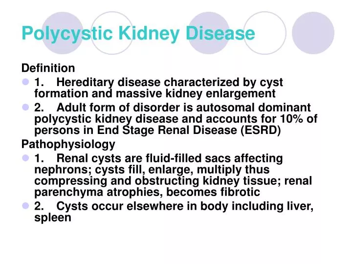
Polycystic Kidney Disease. Definition 1. Hereditary disease characterized by cyst formation and massive kidney enlargement 2. Adult form of disorder is autosomal dominant polycystic kidney disease and accounts for 10% of persons in End Stage Renal Disease (ESRD) Pathophysiology

E N D
Polycystic Kidney Disease Definition • 1. Hereditary disease characterized by cyst formation and massive kidney enlargement • 2. Adult form of disorder is autosomal dominant polycystic kidney disease and accounts for 10% of persons in End Stage Renal Disease (ESRD) Pathophysiology • 1. Renal cysts are fluid-filled sacs affecting nephrons; cysts fill, enlarge, multiply thus compressing and obstructing kidney tissue; renal parenchyma atrophies, becomes fibrotic • 2. Cysts occur elsewhere in body including liver, spleen
Polycystic Kidney Disease Manifestations 1. Disease is slowly progressive; symptoms develop in age 30 – 40’s 2. Common manifestations include • a. Flank pain • b. Microscopic or gross hematuria • c. Proteinuria • d. Polyuria and nocturia (impaired ability to concentrate urine) • e. UTI and renal calculi are common • f. Hypertension from disrupted renal vessels • g. Kidneys become palpable, enlarged, knobby • h. Symptoms of renal insufficiency and chronic renal failure by age of 50 – 60
Polycystic Kidney Disease Collaborative Care: Determine extent of polycystic kidney disease Diagnostic tests • 1. Renal ultrasonography: primary choice for diagnostic; assesses kidney size, identifies and locates renal masses: cysts, tumors, calculi • 2. Intravenous pyelography (IVP): evaluate structure and excretory function of kidneys, ureters, bladder • 3. CT scan of kidneys: detects and differentiates renal masses
Polycystic Kidney Disease Management • 1. Mainly supportive: prevent further renal damage from UTI, nephrotoxic substances, obstruction, hypertension • 2. Fluid intake of 2000 – 2500 mL to prevent UTI, calculi • 3. Control of hypertension with ACE inhibitors and other antihypertensive agents • 4. Eventually require dialysis or transplantation (typically good candidates)
Polycystic Kidney Disease Nursing Care and Nursing Diagnoses • 1. Risk for Ineffective Coping: address genetic counseling and screening for family members • 2. Excess Fluid Volume • 3. Anticipatory Grieving • 4. Knowledge Deficit: of measures to preserve kidney function
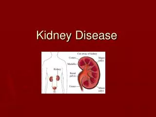
Clients with Renal Failure Definition • 1. Condition in which kidneys are unable to remove accumulated metabolites from blood; leads to altered fluid, electrolyte and acid-base balance • 2. May be due to kidney (primary disorder) or resulting from another disease in another organ or systemic (secondary disorder) • 3. Classified as acute (abrupt onset and may be reversible) or chronic (develops slowly and insidiously with few symptoms until kidneys are severely damaged and unable to meet body’s excretory needs) • 4. Common and costly disease with people with End Stage Renal Disease requiring dialysis or transplant to live • 5. 5 year survival rate for clients on dialysis is 31.3%
Clients with Renal Failure Acute Renal Failure (ARF) • 1. Definition • a. Rapid decline in renal function with azotemia fluid and electrolyte imbalances • b. High mortality rate but is related to clients being seriously ill and aged Risk Factors • a. Major surgery or trauma • b. Infection • c. Hemorrhage • d. Severe heart failure, liver disease • e. Lower urinary tract obstruction • f. Use of nephrotoxic contrast media and medications
Clients with Renal Failure Pathophysiology involved with cause categories a. Prerenal • 1. 55 – 60% cases of ARF • 2. Cause: Conditions that affect renal blood flow and perfusion • A .Decrease vascular volume • B .Decrease cardiac output • C .Decrease vascular resistance b. Intrarenal 1. 35-40% cases of ARF 2. Cause: Acute damage to renal parenchyma and nephron • a. Acute glomerulonephritis • b. Vascular disorders including vasculitis, malignant hypertension, arterial or venous occlusion
Clients with Renal Failure c. Acute Tubular Necrosis (ATN): Destruction of tubular epithelial cell with abrupt decline in renal function from • 1. Prolonged ischemia (>2 hours) as with surgery, severe hypovolemia, sepsis, trauma, burns • 2. Nephrotoxins • a. Aminoglycoside antibiotics • b. Radiologic contrast media • c. Other potential drugs: NSAIDs, heavy metals, ethylene glycol (antifreeze) • 3. Nephrotoxins have increased risk with clients with preexisting renal insufficiency or state of dehydration • 4. Rhabdomyolysis: excess myoglobin from skeletal muscle injury clogs renal tubules (muscle trauma, drug overdose, infection) • 5. Hemolysis: red blood cell destruction
Clients with Renal Failure Postrenal • 1. <5% cases of ARF • 2. Cause: Obstructive; prevents urine excretion • a. Benign prostatic hypertrophy • b. Renal or urinary tract calculi or tumors
Clients with Renal Failure Course and Manifestations of ARF in 3 phases a. Initiation Phase • 1. Lasts hours to day • 2. Begins with initiating event ends when maintenance phase begins • 3. Good prognosis if treated at this phase • 4. Few manifestations; identified when maintenance phase begins b. Maintenance Phase • 1. Characterized by significant fall in GFR and tubular necrosis • 2. Oliguric or non-oliguric but kidneys not eliminating wastes, water, electrolytes, acids: azotemia, fluid retention, electrolyte imbalances (hyperkalemia, hypocalcemia, hyperphosphatemia), acidosis (impaired hydrogen ion elimination) • 3. Anemia after several days due to suppressed erythropoetin secretion; impaired immune function
Clients with Renal Failure • 4. Salt and water retention leading to hypertension and risk for heart failure and pulmonary edema • 5. Hyperkalemia: cardiac dysrhythmias and EKG changes, muscle weakness, nausea, diarrhea • 6. Confusion, disorientation, agitation or lethargy, hyperreflexia, possible seizures, coma • 7. Vomiting, decreased or absent bowel sounds c. Recovery Phase • 1. Progressive tubule cell repair and regeneration; return of GFR to pre-ARF levels • 2. Diuresis occurs as kidney recover but BUN, Creatinine, potassium and phosphate remain high • 3. Renal function improves rapidly first 5 – 25 days but improvement may continue for up to a year
Clients with Renal Failure Collaborative Care a. Prevention of ARF is goal for all clients, especially those at high-risk • 1. Preserve kidney perfusion by adequate vascular volume, cardiac output and blood pressure • 2. Limiting use of nephrotoxic medications or using minimal effective dose, maintaining hydration, monitoring renal function tests b. Treatment goals • 1. Identify and correct underlying cause • 2. Prevent additional renal damage • 3. Restore urine output and kidney function • 4. Compensate for impaired renal function: maintain fluid and electrolyte balance
Clients with Renal Failure Diagnostic tests to identify ARF a. Urinalysis • 1. Fixed specific gravity 1.010 (low) • 2. Proteinuria, if glomerular damage • 3. Presence of red blood cells (glomerular dysfunction), white blood cells (inflammation), renal tubule epithelial cells (ATN) • 4. Cell casts (protein and cellular debris molded in shape of tubular lumen); brown color may indicate hemoglobinuria or myoglobinuria b. Serum BUN and creatinine • 1. Creatinine rises rapidly (24 – 48 hours) and peaks in 5 – 10 days; rise is slower if output maintained • 2. Halt in rise of BUN and Creatinine signals onset of recovery
Clients with Renal Failure Serum Electrolytes • 1. Monitored to determine whether to initiate dialysis • 2. Moderate rise in potassium • 3. Hyponatremia related to water excess d. CBC showed moderate anemia and low hematocrit (Iron and folate may be low and add to anemia) e. Renal ultrasound: used to identify any obstruction, identify acute from chronic renal failure f. CT scan: identify obstruction and kidney size g. IVP, retrograde pyelography, or antegrade pyelography • 1. Assess renal structure and function • 2. Retrograde and antegrade testing less toxicity from contrast media h. Renal biopsy: determine cause, differentiate acute from chronic
Clients with Renal Failure Medications • a. Intravenous fluids and blood volume expanders to restore renal perfusion • b. Low dose Dopamine (Intropin) intravenous infusion to increase renal blood flow and improve cardiac output • c. Diuretic: Furosemide (Lasix) or osmotic diuretic such as mannitol along with intravenous fluids; “washes out” nephrons; prevents oliguria reducing azotemia and electrolyte imbalance • d. Antihypertensive medications including ACE inhibitors to limit renal injury
Clients with Renal Failure Medications to prevent possible complications 1. Prevention of gastrointestinal bleeding (at risk due to stress, impaired platelet function) • a. Antacids • b. H2 receptor antagonists • c. Proton-pump inhibitors 2. Hyperkalemia: serum K > 6.5 mEq/L puts client at risk for cardiac arrest • a. Calcium chlorides • b. Bicarbonate • c. Insulin and glucose
Clients with Renal Failure d. Sodium polystyrene sulfonate (Kayexalete) • 1. Removes potassium from body primarily in large intestine • 2. If given orally, is combined with sorbitol • 3. May be given as retention enema with tap water enema to follow after 30 – 60 minutes 3. Hyperphosphatemia • a. Aluminum hydroxide (AlternaGEL, Amphojel, Nephrox) • b. Binds with phosphates in GI tract and is eliminated from bowel
Clients with Renal Failure Fluid Management • a. Once vascular volume and renal perfusion restored, fluids are restricted • b. Often intake is calculated by adding output from previous 24 hours and 500 ml for insensible losses • c. Fluid balance monitored by daily weights and serum Na level
Clients with Renal Failure Dietary Management • a. Renal insufficiency and underlying disease creates increased rate of catabolism (breakdown of body proteins) and decreased rate of anabolism (tissue repair) • b. Client needs adequate nutrition and calories to prevent catabolism but protein intake needs to be limited to minimize azotemia • c. Protein limited to 0.6g/kg body weight per day; protein should be of high biologic value (contains essential amino acids) • d. Carbohydrate intake is increased for adequate calories and protein-sparing effect
Clients with Renal Failure Dialysis • a. Dialysis is the diffusion of solute molecules across semipermeable membrane from area of higher solute concentration to lower concentration • b. Dialysis used to remove excess fluid, waste products from client with renal failure; can rapidly remove nephrotoxins from blood
Manual peritoneal dialysis via an implanted abdominal catheter (Tenckhoff catheter)
Clients with Renal Failure Hemodialysis: Dialysis process • a. In this type of dialysis, blood is taken from client via vascular access and pumped into a dialyzer; blood is separated from the dialysate (dialysis solution) by semipermeable membrane • b. Processes of diffusion and ultrafiltration remove waste products, electrolytes, excess water • c. Glucose, electrolytes, water can pass through, but larger molecules (protein, red blood cells) are blocked • d. Substances can be added to dialysate to diffuse into the blood of the client • e. Client with ARF may undergo hemodialysis daily initially, then 3 – 4 times/week according to client condition; 3 – 4 hours at a time
Clients with Renal Failure Complications associated with hemodialysis • a. Hypotension, most common, related to changes in osmolality, rapid removal from vascular department, vasodilation • b. Bleeding related to platelet function and use of heparin during dialysis • c. Infection, local or systemic; Staphylococcus aureus septicemia associated with infected vascular access site; higher rates of hepatitis B and C, cytomegalovirus, HIV in hemodialysis clients
Clients with Renal Failure 13. Continuous Renal Replacement Therapy (CRRT) • a. Technique used, which allows more gradual fluid and solute removal than hemodialysis; used for clients with ARF unable to tolerate hemodialysis • b. Done over period of 12 hours or more 14. Vascular Access for Hemodialysis • a. Acute or temporary access is gained inserting double lumen catheter into subclavian, jugular, or femerol vein • b. Blood is drawn from proximal portion of catheter and returned to circulation through distal end of catheter
Clients with Renal Failure Arteriovenous (AV) fistula created for longer term access for dialysis 1. Surgical anastomosis of artery and vein in non-dominant arm, usually radial artery and cephalic vein 2. Usually cannot use fistula for hemodialysis access for a month while it matures 3. Nurse or client can assess functional fistula for complications • a. Thrombosis (clotted off): check for palpable thrill, audible bruit • b. Infection: check for redness, drainage 4. Venipunctures and blood pressures should not be done in arm with the AV fistula 5. AV fistulas are commonly used for vascular access for dialysis clients with chronic renal failure
Clients with Renal Failure Peritoneal dialysis process involves: • a. Peritoneal membrane of client is used as dialyzing surface • b. Warmed sterile dialysate instilled into peritoneal cavity through a catheter that has been inserted into peritoneal cavity • c. Metabolic waster products and excessive electrolytes diffuse into dialysate while it remains in abdomen • d. Water diffusion is controlled by glucose in the dialysate which acts as an osmotic agent • e. Fluid is drained off by gravity into sterile bag at set intervals, thus removing waste products and excess fluid
Clients with Renal Failure Disadvantages of peritoneal dialysis • a. Dialysis is more gradual and may be slow for ARF • b. Risk of peritonitis • c. Contraindicated for clients with abdominal surgery, peritonitis, significant lung disease Health Promotion: Prevention of ARF • a. Maintenance of fluid volume and cardiac output • b. Reduce risk of exposure to nephrotoxins • c. Report output < 30 ml per hour in clients at risk • d. Report dehydration, monitor renal function tests in clients receiving nephrotoxic medications
Clients with Renal Failure Nursing Diagnoses for clients in ARF • a. Excess Fluid Volume • b. Imbalanced Nutrition: Less than body requirements • c. Deficient Knowledge Home care: Client who is recovering from ARF will need teaching for prescribed diet and fluid intake, avoidance of nephrotoxins, prevention of infection, continue under medical supervision
Clients with Renal Failure Client with Chronic Renal Failure (CRF) Definition • a. Progressive renal tissue destruction and loss of function • b. May progress over many years without being recognized until kidneys are unable to excrete metabolic wastes and regulate fluid and electrolytes: End-stage Renal Disease (ESRD) • c. Incidence is increasing especially in older adults; higher in African Americans, Native Americans • d. Conditions causing chronic renal failure diffuse bilateral disease of kidneys with progressive destruction and scarring; diabetes is leading cause of ESRD; then hypertension
Clients with Renal Failure Pathophysiology and Manifestations of Stages a. Decreased Renal Reserve: Early Stage • 1. Unaffected nephrons compensate for lost nephrons • 2. GFR is about 50% of normal • 3. Client is asymptomatic • 4. BUN and serum creatinine are normal b. Renal Insufficiency • 1. GRF falls to 20 – 50% of normal • 2. Azotemia and some manifestations • 3. Insult to kidneys could precipitate onset renal failure (infection, dehydration, exposure to nephrotoxins, urinary tract obstructions)
Clients with Renal Failure c. Renal failure • 1. GRF < 20% of normal • 2. BUN and serum creatinine rise sharply • 3. Oliguria, manifestations of uremia d. End-stage renal disease (ESRD) • 1. GRF < 5 % of normal • 2. Renal replacement therapy necessary to sustain life
Clients with Renal Failure ESRD: Uremia (“urine in blood”) a. Early manifestations • 1. Nausea, apathy, weakness, fatigue • 2. Progresses to frequent vomiting, increasing weakness, lethargy, confusion b. Fluid and electrolyte effects • 1. Urine less concentrated with proteinuria and hematuria • 2. Sodium and water retention • 3. Hyperkalemia (Muscle weakness, paresthesia, EKG changes) • 4. Hyperphosphatemia, hypocalcemia, hypermagesemia • 5. Metabolic acidosis
Clients with Renal Failure Cardiovascular effects • 1. Systemic hypertension • 2. Edema and heart failure; pulmonary edema • 3. Pericarditis: metabolic toxins irritate pericardial sac; less often now with dialysis • 4. Cardiac tamponade: fluid in pericardial sac Hematologic effects • 1. Anemia contributing to fatigue, weakness, depression, impaired cognition, impaired cardiac function • 2. Impaired platelet function Immune system effects • 1. WBC declines • 2. Humoral and cell-mediated immunity impaired • 3. Fever suppressed
Clients with Renal Failure Gastrointestinal effects • 1. Anorexia, nausea, vomiting, hiccups • 2. GI ulcerations, increased risk for GI bleeding • 3. Uremic fetor: urinelike breath odor Neurologic effects • 1. Changes in mentation, poor concentration • 2. Fatigue, insomnia • 3. Psychotic symptoms, seizures, coma • 4. Peripheral neuropathy: “restless leg syndrome”, sensations of crawling, prickling • 5. Muscle weakness, decreased deep tendon reflexes, gait disturbances
Clients with Renal Failure Musculoskeletal effects • 1. Renal osteodystrophy (renal rickets) characterized by osteomalacia (bone softening) and osteoporosis • 2. Bone tenderness and pain Endocrine and metabolic effects • 1. Elevated uric acid levels; risk for gout • 2. Resistance to insulin, glucose intolerance • 3. High triglyceride and < HDL levels resulting in accelerated atherosclerotic process • 4. Menstrual irregularities; reduced testosterone levels Dermatologic effects • 1. Yellowish hue to skin • 2. Dry skin with poor turgor • 3. Pruritis due to metabolic wastes deposited in skin • 4. Uremic frost crystallized deposits of urea on skin
Clients with Renal Failure Collaborative Care • a. Eliminate factors that further decrease renal function • b. Maintenance of nutritional status with minimal toxic waste products • c. Identify and treat complications of CRF • d. Preparation for dialysis or renal transplantation
Clients with Renal Failure Diagnostic Tests: Identify CRF and monitor renal function by following levels of metabolic wastes and electrolytes • a. Urinalysis: fixed specific gravity at 1.010; excess protein, blood cells, cellular casts • b. Urine culture: identify infection • c. BUN and serum creatinine: evaluate kidney function 1. BUN levels • a. Mild azotemia: 20 – 50 mg/dL • b. Severe renal impairment: > 100 mg/dL • c. Uremic symptoms: > 200mg/dL 2. Creatinine levels >4 mg/dL indicate serious renal impairment • d. Creatinine Clearance: evaluates GFR and renal function • 1. Decreased renal reserve: 32.5 – 130 mL/min • 2. Renal insufficiency: 10 – 30 mL/min • 3. ESRD: 5 – 10 mL/min
Clients with Renal Failure • e. Serum electrolytes: monitored throughout course of CRF • f. CBC: moderately severe anemia with hematocrit 20 – 30%; low hemoglobin; reduced RBCs and platelets • g. Renal ultrasonography: CRF: decreased kidney size • h. Kidney biopsy: diagnose underlying disease process; differentiate acute from chronic
Clients with Renal Failure Medications a. General effects of CRF on medication effects • 1. Increased half-life and plasma levels of meds excreted by kidneys • 2. Decreased drug absorption if phosphate-binding agents administered concurrently • 3. Low plasma protein levels can lead to toxicity when protein-bound drugs are given • 4. Avoid nephrotoxic meds or give with extreme caution b. Diuretics (furosemide, other loop diuretics) • 1. Reduce edema • 2. Reduce blood pressure • 3. Lower potassium c. Antihypertensive medications: ACE inhibitors preferred d. Sodium bicarbonate or calcium carbonate correct mild acidosis e. Oral phosphorus binding agents (calcium carbonate, calcium acetate) to lower phosphate levels and normalize calcium levels f. Aluminum hydroxide for acute treatment of hyperphosphatemia
Clients with Renal Failure g. Vitamin D supplements to improve calcium absorption h. To treat dangerously high potassium levels • 1. Intravenous bicarbonate, insulin, glucose • 2. Sodium polystyrene sulfonate (Kayexalate) i. Folic acid, iron supplements to combat anemia j. Multiple vitamin supplement Dietary and Fluid Management • a. Early in course of CRF: diet modifications to slow kidney failure, uremic symptoms, and complications • b. Restrict proteins (40 gm/day) of high biologic value • c. Increase carbohydrate intake (35kcal/kg/day) • d. Limit fluid to 1 – 2 L per day; limit sodium to 2 g/day • e. Restrict potassium (60 -70 mEq/day); no salt substitutes • f. Restrict phosphorus foods (meat, eggs, dairy products)
Clients with Renal Failure Renal Replacement Therapies: considered when medications and dietary modifications are no longer effective • a. Hemodialysis: establish vascular access (create AV fistula) months ahead • b. Peritoneal dialysis: can be initiated when indicated; training client and/or family involved • c. Transplantation: tissue typing and identification of living related potential donors including health assessment of donor Dialysis • a. Considerations • 1. Dialysis manages ESRD, but does not cure it • 2. Hemodialysis or peritoneal dialysis is constant factor of life • 3. Depending on individual client situation and total health, client may prefer death to dialysis
Clients with Renal Failure Hemodialysis for ESRD • 1. Treatments are 3 times per week for 9 – 12 hours • 2. Specific dialysis orders according to body size, residual renal function (based on that day’s current lab test results), dietary intake, concurrent illnesses • 3. Complications during treatment are hypotension and muscle cramps; dialysis disequilibrium syndrome • 4. Long term complications are infection and vascular access problems • 5. Cardiovascular disease is leading cause of death for hemodialysis clients; higher death rate than clients on peritoneal dialysis or transplanted
Clients with Renal Failure Peritoneal Dialysis for ESRD • 1. Continuous ambulatory peritoneal dialysis (CAPD) most common • 2. 2 liters of dialysate instilled into peritoneal cavity and catheter sealed; empty and replace every 4 – 6 hours • 3. Continuous cyclic peritoneal dialysis (CCPD) uses delivery device during nighttime hours and continuous dwell during day Advantages over hemodialysis • a. Eliminates vascular access and heparinization • b. Avoids rapid fluctuation in extracellular fluid • c. Diet intake is more liberal with fluids and nutrients • d. Regular insulin can be added to dialysate to manage hyperglycemia for diabetics • e. Client more able to self-manage