Download
1 / 38
410 likes | 564 Views
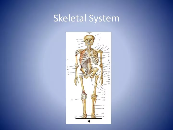
Skeletal System. Introduction. The framework of bones and cartilage that protects our organs and allows us to move is called the skeletal system.

E N D
Introduction • The framework of bones and cartilage that protects our organs and allows us to move is called the skeletal system. • The branch of medicine that deals with the preservation and restoration of the skeletal system, articulations (joints), and associated structures is called orthopaedics.
The skeletal system performs the following functions: • Support • Protection (for internal organs) • Movement • Mineral storage • Storage of blood cell-producing cells • Storage of energy
Bone Composition • Bone is very strong for its relatively light weight • The major components of bone are: • Calcium carbonate • Calcium phosphate • Collagen • Water Cortical Bone Spongy Bone Medullary (marrow) cavity
Bone Composition Cont’d • Calcium carbonate and calcium phosphate: • Make up 60-70% of bone weight • Provide much of the bone’s stiffness and resistance to pressing or squeezing forces • Collagen (a protein): • Gives bone its characteristic flexibility and contributes to its ability to resist pulling and stretching forces • With aging, collagen is lost progressively and bone becomes more brittle. • Water • Bone consists of much smaller proportion of water than other body parts
Bone Classification • According to the degree of porosity, bone can be classified into two general categories: • Cortical bone (low porosity) • Spongy or cancellous bone (high porosity)
Effect of Fitness on Bone • When bones are subjected to regular physical activity and habitual loads, they tend to become denser and more mineralized • e.g. Right forearm of the right-handed tennis player is more dense than her left one from using it more frequently • Inactivity works in the opposite direction, leading to a decrease in weight and strength. • e.g. Loss of bone mass has been noted in bed-ridden patients, inactive senior citizens, and astronauts
Types of Bones • There are five principal types of bones • All bones are classified based on shape
1. Long bones (e.g. thighs, legs, toes, arms, forearms, and fingers) • greater length than width • consist of a shaft and extremities (ends) • slightly curved for strength • consist mostly of compact bone (dense bone with few spaces) but also contain considerable amounts of spongy bone (bone with large spaces)
2. Short bones (e.g. wrist, ankle bones) • Somewhat cube-shaped and nearly equal in length and width • Spongy except at the surface where there is a thin layer of compact bone
3.Flat bones (e.g. cranial bones, sternum, ribs, scapulas) • Generally thin and composed of two more or less parallel plates of compact bone enclosing a layer of spongy bone • Flat bones afford considerable protection and provide extensive areas for muscle attachment
Irregular bones (e.g. vertebrae, and certain facial bones) • Have complex shapes and cannot be grouped into any of the other three categories • They vary in the amount of spongy and compact bone
5.Sesamoid bones • Are small bones in tendons where considerable pressure develops, for instance, the wrist • Their number varies greatly from person to person • All people have at least two sesamoid bones: the patella (kneecap)
Divisions of the Skeletal System • The adult human skeleton consists of 206 bones grouped as the axial skeleton and the appendicular skeleton. • The axial division consists of the bones of the skull, auditory ossicles, hyoid bone, ribs, breastbone, and the backbone. • The appendicular division consists of the bones of the upper and lower extremities (limbs), plus the bones called girdles, which connect the extremities to the axial skeleton. • There are 80 bones in the axial division and 126 in the appendicular. Listed below are the divisions of the skeletal system.
Axial Skeleton Skull Sternum Ribs Vertebral Column
Skull • Divided into two parts: a) Calvaria b) Face
a) Calvaria Parietal Bone Frontal Bone Occipital Bone Temporal Bone
Calvaria Cont. • May be fractured in blows to the skull (e.g., in hockey, being checked and hitting the skull on the ice) • Temporal bone: • more fragile of the calvaria bones • overlies one of the major blood vessels • if fractured and displaced internally = medical emergency (picture)
b) Facial Bones Lacrimal Bone Nasal Bone Zygomatic Bone Maxilla Bone Mandible Bone
Lumbar vertebra, lateral view 7 Cervical Vertebrae (of the neck) 12 Thoracic Vertebrae (of the chest) Lumbar vertebra, superior view 5 Lumbar Vertebrae (of the lower back) Sacrum (mid-line region of buttocks) Coccyx (4 or 5 fused vertebrae of the tail bone) Vertebral Column
Vertebral Column • Vertebrae are arranged in a cylindrical column interspersed with fibrocartilaginous (intervertebral) discs • Function: • provides a strong and flexible support for the body and the ability to keep the body erect • the point of attachment for the muscles of the back. • protect the spinal cord and nerves • absorbs shock through the intervertebral discs without causing damage to other vertebrae
Ribs • Twelve pairs • Made up of : • bone • cartilage which strengthen the chest cage and permit it to expand. • Curved and slightly twisted making it ideal to protect the chest area
Ribs Cont’d • All 12 pairs of ribs articulate with the twelve thoracic vertebrae posteriorly • Classified into three groups based on anterior attachment: (picture) • true ribs • 1-7 • attach to both the vertebrae and the sternum • false ribs • 8-10 • attach only to the sternum indirectly, through 7th rib • floating ribs • 11 and 12 • only attach to the vertebral column
True Ribs (1-7) False Ribs (8-10) Floating Ribs (11-12) The Ribs Manubrium Sternal Body Xiphoid Process Costal Cartilages
Sternum • Mid-line breast bone • The clavicles and ribs one to seven articulate with the sternum Sternum – comprised of the manubrium, sternal body and xiphoid process
Appendicular skeleton Consists of: • 1. The pectoral gridle (chest) • 2. Pelvic girdle (hip) • 3. The upper limbs • 4. The lower limbs
Clavicle Scapula 1.Pectoral Girdle Consists of: • Scapula (shoulder blade) • Clavicle (collar bone) • Allows the upper limb great mobility • The sternoclavicular joint is the only point of attachment between the axial skeleton and the pectoral girdle
2. Pelvic Girdle • Formed by pair of os coxae (hip bones) • supports the bladder and abdominal contents • Attachment: • Posteriorly – join with the sacrum • Anteriorly - join to each other anteriorly • Laterally – join to the head of thigh bone through a cup-shaped acetabulum
Humerus Radius Ulna 3. Upper Limb • Humerus • The arm bone • shoulder to elbow • Radius and Ulna • The forearm bones • elbow to wrist • the radius being located on the thumb side of the hand • when you pronate the forearm, the radius is actually crossing over the ulna - try it yourself
Carpals Proximal Phalanx Metacarpals Phalanges Middle Phalanx Distal Phalanx Upper Limb Cont.
Femur Patella 4. Lower Limb • Femur • thigh bone • from hip to knee • Patella • knee cap • sesamoid bone in the tendon of the quadriceps muscles (thigh)
Fibula Tibia Lat. malleolus Med. malleolus Lower Limb Cont’d • Tibia and Fibula • leg bones • From knee to ankle • Tibia is medial and fibula is lateral • Medial malleolus and Lateral malleolus • The distal ends of the tibia and fibula, respectively • commonly referred to as the "ankle bones" • can be easily palpated
Talus Calcaneus Tarsals Metatarsals Phalanges Lower Limb Cont’d • Tarsals • ankle bones • calcaneus or the heel bone • talus • Metatarsals • 5 bones of the foot • unite with the toes • Phalanges • toe bones • three per toe except the big toe - proximal, middle and distal
Skeletal Surface Markings • The surfaces of bones have various structural features adapted to specific functions. These features are called surface markings. Long bones that bear a great deal of weight have large, rounded ends that can form sturdy joints, for example. Other bones have depressions that receive the rounded ends.