Download
1 / 18
210 likes | 532 Views
RESTRICTIVE THORACIC DISEASE Thoracic Restriction due to causes out with the lungs . Skeletal :Vertebrae-eg Thoracic kyphoscoliosis , Ribs –eg Traumatic multiple rib #s

E N D
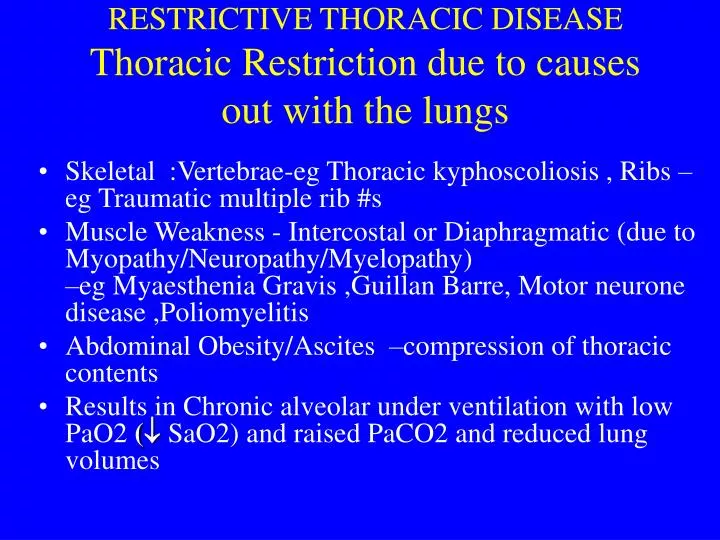
RESTRICTIVE THORACIC DISEASE Thoracic Restriction due to causes out with the lungs • Skeletal :Vertebrae-eg Thoracic kyphoscoliosis , Ribs –eg Traumatic multiple rib #s • Muscle Weakness - Intercostal or Diaphragmatic (due to Myopathy/Neuropathy/Myelopathy) –eg Myaesthenia Gravis ,Guillan Barre, Motor neurone disease ,Poliomyelitis • Abdominal Obesity/Ascites –compression of thoracic contents • Results in Chronic alveolar under ventilation with low PaO2 ( SaO2) and raised PaCO2 and reduced lung volumes
RESTRICTIVE THORACIC DISEASE • Due to disease within the lungs –ie Interstitial Lung Disease • Disease of alveolar structures • - alveolar walls/lumen ( lung interstitium ) • Pathophysiology • impaired alveolar gas exchange • - alveolar barrier to O2 exchange • (ie Alveolar-Arteriolar barrier ) • - CO2 exchange unimpaired as alveolar ventilation normal (CO2 v soluble and blown off ) • : PaO2 ( SaO2) normal PaCO2
Aetiology of ILD • Fluid in the alveolar air spaces • Cardiac Po oedema (in alv walls and lumen) due to raised Po venous pressure –ie LVF • Non Cardiac Po oedema –Normal Po venous pressure with leaky Po capillaries -due to sepsis or trauma (Shock lung or ARDS)-due to Altitude sickness
AETIOLGY OF ILD Consolidation of alveolar air spaces: • Infective pneumonia - viral, bacterial, fungal, protozoal • Infarction - pulmonary emboli/vasculitis • Other causes (ie BOOP) - rheumatoid disease - drugs - cryptogenic
AETIOLGY OF ILD Inflam Infiltrate of alveolar walls (ie Alveolitis): •Granulomatous-alveolitis •Extrinsic-Allergic-Alveolitis (Hypersensitivity Pneumonitis-Type 3 reaction) - Farmers lung - Avian (pigeon, budgie) • Sarcoidosis -Multisystem disease -Lymphadenopathy/Erythema nodosum Uveitis/Myocarditis/Neuropathy
Aetiology of ILD Alveolitis continued • Drug induced alveolitis - Amiodarone - Bleomycin, Methotrexate - Gold • Fibrosing alveolitis - Rheumatoid , Cryptogenic • Autoimmune(multisystem) -SLE, Polyarteritis,Wegeners,Churg-Strauss
AETIOLGY OF ILD Dust-disease (Pneumoconiosis) • Pulmonary fibrosis - asbestosis - silicosis
AETIOLGY OF ILD Carcinomatosis • Lymphatic (adenoca) -bronchus,breast,prostate,colon,stomach Eosinophilic • Drugs - Nitrofurantoin • Fungal - Aspergillosis • Parasites - Ascaris, Filariasis • Autoimmune vasculitis -Churg-Strauss,Polyarteritis
CLINICAL SYNDROME OF ILD • Breathless on exertion • No cough or wheeze • Lung crackles (inspiratory) • Finger clubbing • Central cyanosis (if hypoxaemic) • Pulmonary fibrosis(honeycomb lung) • End stage response to any inflammatory process
DIAGNOSIS OF ILD #1 • History-eg occupation,drugs,pets,arthritis • Reduced lung volumes • : FEV1 FVC1 normal ratio > 75% • : Peak flow normal • Reduced gas diffusion (TLCO) • Arterial oxygen desaturation (PaO2 SaO2) • - at rest or on exercise
DIAGNOSIS OF ILD #2 • Antibodies:Avian,Fungal,Auto-antibodies (Rheumatoid,Antinuclear) • Serum ACE and Ca raised in Sarcoid • Bilateral diffuse alveolar infiltrates on chest X-ray • Echocardiogram to excl LVF • High resolution CT scan-Inflammatory ground glass vs Fibrotic nodular components of alveolar infiltrates • Transbronchial or thoracoscopic lung biopsy -rarely indicated
TREATMENT OF ILD • Remove any trigger factor • - dust, drug, allergen • Treat any inflammation-immunosuppressives • 1st line Prednisolone • 2nd line • Azathioprine Cyclophosphamide • Cylcosporin • O2 if hypoxaemic
Bilateral hilar lymphadenopathy and lung infiltrares -Sarcoidosis