Download
1 / 63
650 likes | 870 Views
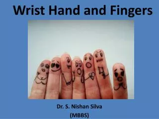
Wrist, Hand, Elbow & Shoulder. Chapters 12, 11 & 10. Anatomy of the Wrist and Hand. Looks a lot like the foot Has similar bone structures: Phalanges Metatarsals Carpals There are 26 bones There are many ligaments that hold the structure of the hand together. Carpal Bones. Proximal:

E N D
Wrist, Hand, Elbow & Shoulder Chapters 12, 11 & 10
Anatomy of the Wrist and Hand • Looks a lot like the foot • Has similar bone structures: • Phalanges • Metatarsals • Carpals • There are 26 bones • There are many ligaments that hold the structure of the hand together
Carpal Bones Proximal: A=Scaphoid B=Lunate C=Triquetral D=Pisiform Distal: E=Trapezium F=Trapezoid G=Capitate H=Hamate
The Scaphoid Bone • Find your anatomical snuff box • Only blood supply at one end of the bone • It has difficulty healing if the blood supply is interrupted
Joints of the wrist and hand • There are three phalanges in each finger and two in the thumb • Distal, middle and proximal • Joints: • Distal Interphalangeal jnt (DIP) • Proximal Iinterphalangeal jnt (PIP) • Metacarpal Phalangeal jnt (MCP) • Carpometacarpal jnt (CMP)
Muscles of the hand and forearm • There are two major groups of muscles at the wrist and forearm • Flexors: on the dorsal side of the hand • Extensors: on the ventral side of the hand
The Thumb • Testing the ulnar collateral ligament of the thumb • The collateral ligaments of the thumb provide the majority of its stability
Preventing injuries to the hand Boxing Batting Cycling Field hockey/ girls lacrosse
More Gloves Lineman Gloves Receiver/ Running back gloves
Wrist Sprains • Occur from twisting and overuse • Injured structure depends upon the stress placed on the wrist • Ulnar Deviation is movement towards the ulnar
The Lunate • Dislocation of the lunate bone occurs more often than any other carpal dislocation • Presents as deformity, pain, swelling, and decreased range of motion
Ganglion Cyst • A pocket of fluid within the sheath • Should be referred to a physician • Sometimes is removed surgically
Gamekeepers/Skiers Thumb • Thumb is forced into abduction forcefully • Pain over the joint, swelling may be present • An x-ray may be necessary to rule out a fracture
A “Jammed” Finger • A sprain of the collateral ligaments in the finger
Finger Dislocations Don’t JUST “pop” it !!!!! • There could be underlying hidden issues going on at the joint • There could be a tendon rupture or a fracture!!!
Fractures • Boxer’s fracture is most common in athletes for many reasons
Fractures • Other fractures require the same care and treatment
Muscle and Tendon Injuries • Repetitive stress and stretching can cause injuries to these structures • Some of these include • Carpal tunnel • deQuervian’s tendinitis • Mallet Finger • Jersey finger • Boutonniere deformity
Carpal tunnel • Most common as an overuse injury • Tennis • Field hockey • Watch for acute carpal tunnel due to poor position in slings and casts
deQuervian’s tendinitis • Abductor pollicis longus & Extensor Pollicis brevis tendons • Prolonged or repetitive radial deviation (shot putters) • Swelling, crepitus and pain with abduction
Mallet Finger • An avulsion fracture of the distal phalanx. • Cannot extend the distal phalanx
Jersey finger • Avulsion fracture of flexor tendon • Unable to flex the DIP
Boutonniere deformity • Deformity arises when there is a rupture of the central slip of the extensor mechanism. • This is an uncommon sporting injury usually due to an end-on injury to the finger with sudden bending at the P.I.P. joint • Often in football or basketball
Elbow • This is a very bony joint • Common to have contusions all around the elbow. • Use PRICES
Ligaments • There is thick joint capsule surrounding the elbow. • Relies on the ligaments for stability • Ulnar collateral • Radial collateral • Annular
Muscles • Biceps- elbow flexion • Triceps- elbow extension • Wrist flexors- medial epicondyle of humerus • Wrist extensors- lateral epicondyle of humerus
Preventing Injuries to the Elbow • Not a frequently injured joint • Many of the injuries are caused by overuse • Most injuries occur in racket sports such as tennis, or overhead throwing sports such as baseball and softball. • Many times injuries are brought about by poor training
Preventing Injuries to the Elbow • Athletes train the “beach muscles” • Overwork the Biceps to get “ pipes” or “guns” • What about the Triceps??? • What about the wrist flexors and extensors??
What about equipment? • Tennis players can cause themselves injuries if the grip is too small on the racket. • Throwers should have a strong tricep and lots of flexibility in the elbow to prevent injuries.
Ulnar collateral More common in throwers The stress of overhead activity strains the medial aspect of the elbow. Wrestling? Pain and swelling treat as any other ligament sprain Radial Collateral These are rare Treat the same as a ulnar collateral sprain. Sprains
Vascularity and Nerves • There are numerous blood vessels and three major nerves that pass though t he elbow
Epicondylitis Lateral Epicondylitis • aka: Tennis Elbow • Poor mechanics and overuse • Presents as pain and swelling at the lateral epicondyle • Treat with PRICES Medial Epicondylitis aka: Little League Elbow Repetitive Throwing Little league elbow may have an avulsion fracture at the epiphysis
Fractures • Elbow fractures are rare in athletics. • Often results from a forceful blow to the area or landing on hard surface.
Elbow Dislocation • One of the most commonly dislocated joints in the body • Doesn’t take a lot of force to dislocate the joint • MUST BE SEEN by MD immediately
Olecranon Bursitis • PRICES • Use a compression wrap or sleeve to alleviate swelling • May have to be drained by MD • Not always painful
Shoulder Anatomy A separation occurs here at the acromio-clavicular joint A dislocation occurs here at the gleno-humeral joint
Bony Anatomy • Three bones: • Humerus • Bicipital groove • Clavicle • S shape • Scapula • Corocoid process • Acromion process (a/c joint) • Many ligaments • Not a very stable joint
There are many joints at the shoulder Most commonly injured joints are Acromio-clavicular Gleno-humeral Each held together by many ligaments JOINTS
Muscles of the shoulder • The Rotator Cuff • SITS muscles • Supraspinatus • Infraspiantus • Teres minor • Subscapularis • Deltoid • Lays over the head of the humerus • Pectoralis • Originate at sternum attach to the anterior portion of the humerus
Biceps- two heads Originates at the Coracoid process and the Humerus Distal attachment is a the radial head Runs through the bicipital groove ACTION: elbow flexion and forearm supination Triceps Originates at the posterior humeral head and scapula Distal attachment is distal humerus at the elbow ACTION: Elbow extension and shoulder extension Muscles