Download
1 / 24
240 likes | 247 Views
Molecules of Life. Chapter 3 Part 2. 3.5 Proteins – Diversity in Structure and Function. Proteins are the most diverse biological molecule (structural, nutritious, enzyme, transport, communication, and defense proteins). Proteins and Amino Acids. Protein

E N D
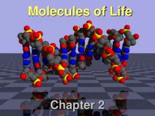
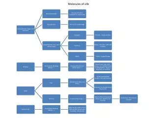
Molecules of Life Chapter 3 Part 2
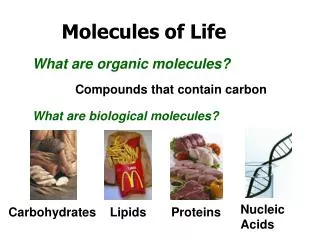
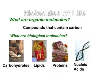
3.5 Proteins – Diversity in Structure and Function • Proteins are the most diverse biological molecule (structural, nutritious, enzyme, transport, communication, and defense proteins)
Proteins and Amino Acids • Protein • An organic compound composed of one or more chains of amino acids
Polypeptides • Protein synthesis involves the formation of amino acid chains called polypeptides • Polypeptide • A chain of amino acids bonded together by peptide bonds in a condensation reaction between the amine group of one amino acid and the carboxyl group of another amino acid
A DNA encodes the order of amino acids in a new polypeptide chain. Methionine (met) is typically the first amino acid. B In a condensation reaction, a peptide bond forms between the methionine and the next amino acid, alanine (ala) in this example. Leucine (leu) will be next. Stepped Art Fig. 3-16a, p. 44
CA peptide bond forms between the alanine and leucine. D The sequence of amino acid subunits in this newly forming peptide chain is now met–ala–leu–trp. The process may continue until there are hundreds or thousands of amino acids in the chain. Tryptophan (trp) will be next. The chain is starting to twist and fold as atoms swivel around some bonds and attract or repel their neighbors. Stepped Art Fig. 3-16b, p. 45
Levels of Protein Structure • Primary structure • The sequence of amino acids in a protein • Secondary structure • The polypeptide chain folds and forms hydrogen bonds between amino acids
Levels of Protein Structure • Tertiary structure • A secondary structure is compacted into structurally stable units called domains • Forms a functional protein • Quaternary structure • Some proteins consist of two or more folded polypeptide chains in close association
a) Protein primary structure: Amino acids bonded as a polypeptide chain. b) Protein secondary structure: A coiled (helical) or sheetlike array held in place by hydrogen bonds (dotted lines) between different parts of the polypeptide chain. helix (coil) sheet c) Protein tertiary structure: A chain’s coils, sheets, or both fold and twist into stable, functional domains such as barrels or pockets. barrel d) Protein quaternary structure: two or more polypeptide chains associated as one molecule. Stepped Art Fig. 3-17, p. 45
Just One Wrong Amino Acid… • Hemoglobin contains four globin chains, each with an iron-containing heme group that binds oxygen and carries it to body cells • In sickle cell anemia, a DNA mutation changes a single amino acid (glutamate to valine) in a beta chain, which changes the shape of the hemoglobin molecule, causing it to clump and deform red blood cells
glutamic acid glutamic acid valine histidine leucine threonine proline A Normal amino acid sequence at the start of the hemoglobin beta chain. Fig. 3-19a, p. 47
valine histidine leucine threonine proline valine glutamic acid B One amino acid substitution results in the abnormal beta chain of HbS molecules. The sixth amino acid in such chains is valine, not glutamic acid. Fig. 3-19b, p. 47
Proteins Undone – Denaturation • Heat, changes in pH, and salts can disrupt the hydrogen bonds that maintain a protein’s shape • When a protein loses its shape and no longer functions, it is denatured • Ex. Albumin (egg white)
3.7 Nucleic Acids • Some nucleotides are subunits (monomers) of nucleic acids (polymers) ; DNA and RNA
Nucleotides • Nucleotide • A small organic molecule consisting of a sugar with a five-carbon ring, a nitrogen-containing base, and one or more phosphate groups • ATP (Energy Currency of the Cell) • A nucleotide with three phosphate groups • Important in phosphate-group (energy) transfer
Nucleic Acids • Nucleic acids • Polymers of nucleotides in which the sugar of one nucleotide is attached to the phosphate group of the next • RNA and DNA are nucleic acids
RNA • RNA (ribonucleic acid) • Contains four kinds of nucleotide bases, including ATP • Important in protein synthesis
DNA • DNA (deoxyribonucleic acid) • Two chains of nucleotides twisted together into a double helix and held by hydrogen bonds • Contains all inherited information necessary to build an organism
covalent bonding in sugar– phosphate backbone hydrogen bonding between bases Fig. 3-22, p. 49