Download
1 / 33
330 likes | 335 Views
General Toxicology Toxic Responses of the R espiratory System (I) Lec.5 4 th Year 2018-2019 University of Mustansiriyah/College of Pharmacy Department of Pharmacology & Toxicology Lecturer Rua Abbas Al-Hamdy. Objectives of this lecture are to:

E N D
General ToxicologyToxic Responses of the Respiratory System (I)Lec.54th Year2018-2019University of Mustansiriyah/College of PharmacyDepartment of Pharmacology & ToxicologyLecturer Rua Abbas Al-Hamdy
Objectives of this lecture are to: • determine the respiratory tract structure & function. • determine acute & chronic responses of the lung to injury.
Overview of the respiratory system: • The respiratory system has direct contact with the inhaled air. • Inhaled air contains a variety of environmental pollutant (e.g., gas, dust, fiber & tobacco smoke). • Air also contains air borne viruses, bacteria & fungi. • The respiratory system is also exposed to inhaled drugs which are used for local or systemic effect.
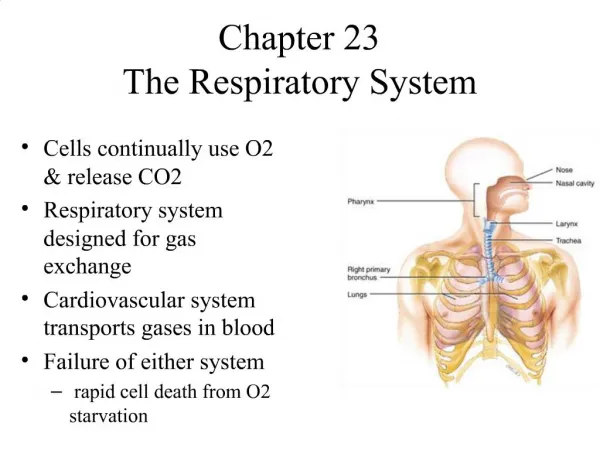
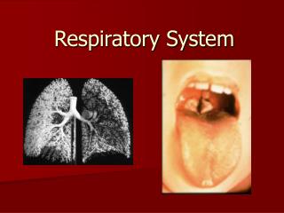
Respiratory tract structure & function: • I. Oronasal passages: • The respiratory tract is divided into the upper respiratory tract (from the nostril or mouth to the pharynx) & lower respiratory track (airway passages & lung parenchyma below the pharynx) (Fig. 1). • The upper respiratory track functions to conduct, heat, humidify, filter, & chemosense incoming air. • Nasal epithelia can metabolize many foreign compounds by cytochrome P450 & other enzymes.
The olfactory epithelium contains specialized chemosensory olfactory neurons located in the superior portion of the nasal passage. • The main nerve endings that perceive irritants, the chemical nociceptors also discern temperature & mechanical stress. Two protein families, the transient receptor potential (TRP) channels & the taste (TAS) receptors, perform these functions in the upper respiratory tract.
TRP channels are ion channels that are permeable to cations, including calcium, magnesium, & sodium. • Among the subfamilies of TRP receptors, are TRP subfamily A (TRPA) & TRP subfamily V (‘’V” for vanilloid) (TRPV). TRPA1 & TRPV1 are the major irritant receptors in the nasal passage & are primarily within the trigeminal nerve.
II. Conducting airways: • The conducting airways of the lower respiratory tract can be divided into: • proximal (trachea & bronchi), & • distal regions (bronchioles). • The epithelium of the proximal airway & a portion of the nasal passage has specialized cells. These cells include ciliated, mucous& basal cells. • These cells work together to form a mucous layer that traps & removes inhaled material via mucociliary clearance.
In humans, the bronchiolar secretoglobin cells (BSCs), previously called the Clara cell, are found mainly in the distal airways. BSCs are known to inhibit phospholipase A2 & limit inflammation.
III. Gas exchange region: • The gas exchange region consists of terminal bronchioles, respiratory bronchioles, alveolar ducts, alveoli, blood vessels, & lung interstitium. Gas exchange occurs in the alveoli. • The alveolar epithelium consists of two cells, the alveolar type I & type II cell. • Alveolar type I cells cover 95% of the alveolar surface, & have a role in gas exchange.
Functions of alveolar type II cells: • They produce & secrete suractant, a mixture of lipids, & four suractant associated proteins. • They can undergo mitotic division & replace damaged type I cells. • Particles deposited in the alveolar region are removed by specialized cells, the alveolar macrophage.
Responses of the lung to injury: • Acute responses, & • Chronic responses.
Acute responses of the lung to injury: • Trigeminally mediated airway reflexes. • Bronchoconstriction, airway hyperreactivity, & neurogenic inflammation. • Acute lung injury (pulmonary edema).
Trigeminally mediated airway reflexes: • Nasal & airway irritation represents a common response to inspired toxic compounds. • Nasal irritation is mediated by irritant receptors [eg, transient receptor potential cation channel-A1(TRPA1)] that trigger trigeminal nerves characterized by tickling, itching & painful nasal sensations. • TRPA1 is sensitive to several irritants including acrolein, allylisothiocyanate, chlorine, & hydrogen peroxide.
If continued exposure cannot be avoided, many irritants will produce cell necrosis.
Bronchoconstriction, airway hyperreactivity & neurogenic inflammation: • Bronchoconstriction can be provoked by: • irritants (e.g., acrolein) • cigarette smoke, • air pollutants, • cholinomimetic drugs (acetylcholine), • histamine, • prostaglandins (PGs) (mainly PGF2α & PGD2), & • leukotrienes.
Characteristic symptoms include wheezing, coughing, a sensation of chest tightness & dyspnea. • Irritants can prime the autonomic response by lowering the threshold dose of acetylcholine needed to induce bronchoconstriction. A lower threshold of acetylcholine-mediated bronchoconstriction is called airway hyperreactivity (or hyperresponsiveness). • Irritants can also stimulate TRP channels that cause neurogenic inflammation.
Acute lung injury (pulmonary edema): • Acute lung injury (both adult or infant respiratory distress syndrome) is marked by: • alveolar epithelial & endothelial cell damage, & • inflammatory cell influx. • These events lead to surfactant disruption & pulmonary edema. • Pulmonary edema produces a thickening of the alveolar capillary barrier & thereby limits O2 & CO2 exchange.
During acute lung injury, profibrotic growth factors, transforming growth factor beta 1 (TGFB1) is activated. • When inhaled in high concentrations, acrolein, HCl, NO2, NH3, or phosgene may produce immediate alveolar damage leading to a rapid death. • However, these gases inhaled in lower concentrations may produce very little apparent damage in the respiratory tract.
Chronic responses of the lung to injury: • Chronic obstructive pulmonary disease (COPD) • Lung cancer • Asthma • Pulmonary fibrosis
Chronic obstructive pulmonary disease (COPD): • COPD involves an airway (bronchitis) & an alveolar (emphysema) pathology. • Chronic bronchitis is defined by the presence of sputum production & cough for at least three months in each of two consecutive years. • In emphysema, destruction of the gas-exchanging surface area results in a distended, hyperinflated lung that no longer effectively exchanges oxygen & carbon.
Lung cancer: • The increased risk of developing lung cancer for average smokers compared with nonsmokers is 8- to 10-fold & for heavy smokers about 20- to 40-fold. • The main factor responsible for smoking dependence is nicotine. • The global burden is ∼1.2 million cancer deaths per year, with 85% of lung cancer cases in men & 47% of lung cancer cases in women being attributable to tobacco use.
Arsenic, asbestos, beryllium, cadmium, chromium, & nickel have been associated with cancer of the respiratory tract.
Asthma: • Asthma is characterized clinically by attacks of shortness of breath, which is caused by narrowing of the large conducting airways (bronchi). • In allergic asthma, previous exposure to an antigen typically leads to the generation of immunoglobin E (IgE).
Upon reexposure, the antigen causes: • cross-linking of IgE molecules • activation of lymphocytes, eosinophils, macrophages, & mast cells • with elaboration & release of an array of cytokines, eicosanoids, histamine, tachykinins & other mediators. • Asthma has been associated with a number of occupations. • Occupational asthma can be induced by high-molecular-weight & some low-molecular-weight substances.
High-molecular-weight agents including flour-, cereals-, latex- or animal-derived proteins & enzymes cause sensitization through an IgE-mediated mechanism, such as in common atopic asthma. • Many low molecular-weight agents that induce occupational asthma include acid anhydrides & platinum salts that induce asthma through an IgE mechanism, but most low-molecular-weight agents involve an uncertain mechanism of induction.
Many of the low-molecular-weight agents can cross-link biological macromolecules. These agents include: • metals (e.g., nickel, chromium, cobalt, zinc, cadmium, & aluminum), • diisocyanates, • cleaning agents, • wood dusts, & • pesticides.
Pulmonary fibrosis: • The pathological hallmark of pulmonary fibrosis is increased focal staining of collagen fibers in the alveolar interstitium. • Excess lung collagen is usually observed not only in the alveolar interstitium but also throughout the centriacinar region, including the alveolar ducts & respiratory bronchioles (Fig. 2). • The pleural surface of the lung may also become fibrotic & together with parenchymal stiffening prevent full lung inflation.
A Figure 2. Normal rat lung & pulmonary fibrosis induced by asbestos in rat lung. A: Normal rat lung. B: Lung of a rat one year after exposure to chrysotile asbestos. Note accumulation of connective tissue around blood vessel & airways (fibrosis). B
The pathogenesis of pulmonary fibrosis involves epithelial cell injury & macrophage activation produced by a wide range of toxic insults. For example, macrophages can be activated by phagocytosis of crystalline silica which activates inflammasome receptor-mediated tumor necrosis factor (TNF) & interleukin 1 beta (IL1B) formation. • Epithelial cells & macrophages also release chemokines that recruit & activate other inflammatory cells including neutrophils & T cells.
These cells combine to produce excessive TGFB1, TNF, IL1B, IL13, and IL17. Of these, TNF & TGFB1 are major mediators in pulmonary fibrosis. • The consequence of the combined TGFB1/3, cytokine release, & hypoxia leads to epithelial-to-mesenchymal transition in which lung epithelial cells transdifferentiate into fibroblast-like cells. These cells contribute to a larger population of myofibroblasts that arise from local mesenchymal cells, & bone marrow–derived fibrocytes.
The migration, proliferation, & activation of myofibroblasts contribute to excessive excellular matrix deposition that has altered collagen cross-linking. The consequence of this process is disorderly repair & substained fibrogenesis, which leads to progressive stiffness of the fibrotic lung.