Download
1 / 32
460 likes | 1.74k Views
Breathing exercises Mazyad Alotaibi. Respiration Respiration can be divided into :. External respiration : ventilation and exchange of gasses in the lungs. Internal respiration : ventilation and exchange of gasses in the tissues. Exchange occurs as result :

E N D
RespirationRespiration can be divided into: • External respiration: ventilation and exchange of gasses in the lungs. • Internal respiration: ventilation and exchange of gasses in the tissues. Exchange occurs as result: - Ventilation: mechanical - Diffusion: random movement
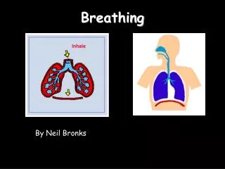
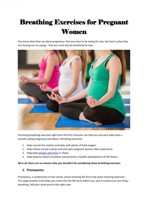
Breathing • Breathing (pulmonary ventilation). consists of two cyclic phases: • Inhalation, also called inspiration - draws gases into the lungs. • Exhalation, also called expiration - forces gases out of the lungs.
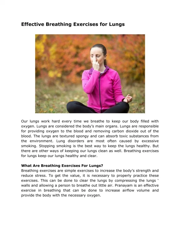
Respiratory System Functions • supplies the body with oxygen and disposes of carbon dioxide • filters inspired air • produces sound • contains receptors for smell • helps regulate blood PH
Structures of the Respiratory System • Consists of an upper respiratory tract (nose to larynx) and a lower respiratory tract ( trachea onwards) . • Conducting portion transports air. - includes the nose, nasal cavity, pharynx, larynx, trachea, and progressively smaller airways, from the primary bronchi to the terminal bronchioles • Respiratory portion carries out gas exchange. - composed of small airways called respiratory bronchioles and alveolar ducts as well as air sacs called alveoli
Upper Respiratory Tract • Composed of the nose and nasal cavity, paranasal sinuses, pharynx (throat), larynx. • All part of the conducting portion of the respiratory system.
Lower Respiratory Tract • Conducting airways (trachea, bronchi, up to terminal bronchioles). • Respiratory portion of the respiratory system (respiratory bronchioles, alveolar ducts, and alveoli).
Muscles of Inspiration • The primary muscle of inspiration is - Diaphragm which : works during quiet breathing forces abdominal contents down & forward • Accessory muscles which work during strenuous physical activities are: External intercostals, pectoralis minor, scaleni & sternocleidomastoid : they lift ribs up and outwards
Muscles of Expiration • Expiration during quiet breathing is passive due to elastic recoil of chest cavity • Decrease in lung size force air out of lungs • During exercise and voluntary hyperventilation, • rectus abdominus, transverse abdominus: push diaphragm up • internal intercostals: pull ribs downwards
Primary and AccessoryVentilatory Muscles Inspiration • Primary muscles: diaphragm, scalenes, parasternals •Accessory muscles: sternocleidomastoids, upper trapezius, pectoralis major and minor, subclavius, and possibly the external intercostals Expiration • Primary muscles: none active during tidal (resting) expiration •Accessory muscles: abdominals including the rectus abdominis, transversus abdominis, and internal and external obliques; pectoralis major; and possibly the internal intercostals
Goals of Breathing Exercises • Improve or redistribute ventilation. • Increase the effectiveness of the cough mechanism and promote airway clearance. • Prevent postoperative pulmonary complications. • Improve the strength, endurance, and coordination of the muscles of ventilation. • Maintain or improve chest and thoracic spine mobility. • Correct inefficient or abnormal breathing patterns and decrease the work of breathing. • Promote relaxation and relieve stress. • Teach the patient how to deal with episodes of dyspnea. • Improve a patient’s overall functional capacity for daily living, occupational, and recreational activities
Examination Evaluation of the patient with pulmonary dysfunction and determination of a diagnosis, prognosis, and intervention plan are based on the findings derived from a comprehensive examination, including a history, systems review, and specific tests
Components of the Examination General Appearance of the Patient Analysis of Chest Shape and Dimensions Symmetry of the chest and trunk. Observe anteriorly, posteriorly, and laterally; the thoracic cage should be symmetrical. Mobility of the trunk. Check active movements in all directions and identify any restricted spinal motions, particularly in the thoracic spine. Shape and dimensions of the chest. The anteroposterior(AP) and lateral dimensions are usually 1:2. Posture or Preferred Positioning Breathing Pattern Assess the rate, regularity, and location of ventilation at rest and with activity. A normal respiratory rate for a healthy adult is 12 to 20 breaths per minute. Chest Mobility Symmetry of chest movement. Analysis of the symmetry of the moving chest during breathing gives the therapist information about the mobility of the thorax and indicates indirectly what areas of the lungs may or may not be responding.
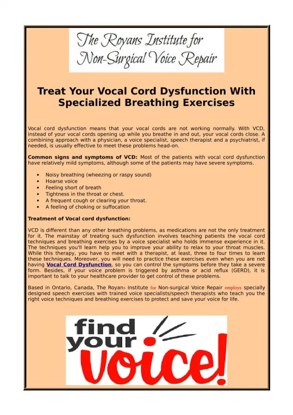
Abnormal Breathing Patterns • Dyspnea. Distressed, labored breathing as the result of shortness of breath. Tachypnea. Rapid, shallow breathing; decreased tidal volume but increased rate; associated with restrictive or obstructive lung disease and use of accessory muscles of inspiration. Bradypnea. Slow rate with shallow or normal depth and regular rhythm; may be associated with drug overdose. Hyperventilation. Deep, rapid respiration; increased tidal volume and increased rate of respiration; regular rhythm. Orthopnea. Difficulty breathing in the supine position. Apnea. Cessation of breathing in the expiratory phase. Apneusis. Cessation of breathing in the inspiratory phase.
Principles of breathing exercises If possible, choose a quiet area for instruction in which therapist can interact with the patient with minimal distractions. Explain to the patient the aims and rationale of breathing exercises Have the patient assume a comfortable, relaxed position Observe and assess the patient’s spontaneous breathing pattern while at rest and later with activity. Demonstrate the desired breathing pattern to the patient. Have the patient practice the correct breathing pattern in a variety of positions at rest and with activity.
Precautions: When teaching breathing exercises, be aware of the following Never allow a patient to force expiration. Expiration should be relaxed or lightly controlled. Do not allow a patient to take a highly prolonged expiration. Do not allow the patient to initiate inspiration with the accessory muscles and the upper chest. Allow the patient to perform deep breathing for only three or four inspirations and expirations at a time to avoid hyperventilation.
Breathing patterns and procedures Diaphragmatic breathing The patient places his or her own hands on the abdomen to feel the movement of proper diaphragmatic breathing. By placing the hands on the abdomen, the patient can also feel the contraction of the abdominals, which occurs with controlled expiration or coughing
Segmental Breathing - Are designed to expand localized areas of the lung Two examples of segmental breathing that target the lateral and posterior segments of the lower lobes are described. However, segmental breathing techniques also may need to be directed to the middle and upper lobes if there is accumulation of secretions or insufficient lung expansion in these areas. Lateral Costal Expansion: can be carried out unilaterally or bilaterally Deep breathing while focusing on movement of the lower portion of the rib cage may facilitate diaphragmatic excursion. This technique is particularly important for the patient with a stiff lower rib cage, as is often seen with chronic bronchitis, emphysema, or asthma.
Bilateral lateral costal expansion—supine. Bilateral lateral costal expansion sitting
The patient applies his or her own manual pressure during lateral costal expansion.
Belt exercises reinforce lateral costal breathing (A) by applying resistance during inspiration and (B) by assisting with pressure along the rib cage during expiration.
Posterior Basal Expansion Deep breathing emphasizing posterior basal expansion is important for the postsurgical patient who is confined to bed in a semireclining position for an extended period of time because secretions often accumulate in the posterior segments of the lower lobes. Procedure Have the patient sit and lean forward on a pillow, slightly bending the hips . The therapist hands is placed over the posterior aspect of the lower ribs, and follow the same procedure just described for lateral costal expansion.
Posterior basal expansion: the therapist hands is placed over the posterior aspect of the lower ribs. Right middle lobe expansion: in which the therapist hands at either the right or the left side of the patient’s chest, just below the axilla. Apical expansion: in which pressure is applied below the clavicle with the fingertips
Pursed Lip Breathing: Pursed-lip breathing is a strategy that involves lightly pursing the lips together during controlled exhalation. This breathing pattern often is adopted spontaneously by patients with COPD to deal with episodes of dyspnea. Patients with COPD using pursed-lip breathing report a decrease in their perceived level of exertion during activity. Procedure Have the patient assume a comfortable position and relax as much as possible. Have the patient breathe in slowly and deeply through the nose and then breathe out gently through lightly pursed lips as if blowing on and bending the flame of a candle but not blowing it out. Explain to the patient that expiration must be relaxed and that contraction of the abdominals must be avoided. Place your hand over the patient’s abdominal muscles to detect any contraction of the abdominals.
EXERCISES TO MOBILIZE THE CHEST Chest mobilization exercises are any exercises that combine active movements of the trunk or extremities with deep breathing. They are designed to maintain or improve mobility of the chest wall, trunk, and shoulder girdles when it affects ventilation or postural alignment. For example, a patient with hypomobility of the trunk muscles on one side of the body does not expand that part of the chest fully during inspiration. Exercises that combine stretching of these muscles with deep breathing improve ventilation on that side of the chest.
Chest mobilization during inspiration and expiration.To mobilize the lateral rib cage have the patient (A) bend awayfrom the tight side during inspiration and (B) bend toward the tightside during expiration.
(A) A stretch is applied to the pectoralis muscles during inspiration, and (B) the patient brings the elbows together to facilitate expiration.
(A) Chest expansion is increased with bilateral movement of the arms overhead during inspiration. (B) Expiration is then reinforced by reaching the arms toward the floor.