Download
1 / 1
10 likes | 153 Views
Integrating CT in Minimally Invasive Treatment of the Coronary Arteries. D Ruijters, N H Bakker, O Wink, M Vembar, G Lavi, B M ter Haar Romeny, P Suetens Philips Medical Systems, the Netherlands danny.ruijters@philips.com, onno.wink@uchsc.edu. Abstract

E N D
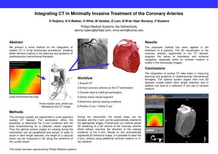
Integrating CT in Minimally Invasive Treatment of the Coronary Arteries D Ruijters, N H Bakker, O Wink, M Vembar, G Lavi, B M ter Haar Romeny, P Suetens Philips Medical Systems, the Netherlandsdanny.ruijters@philips.com, onno.wink@uchsc.edu Abstract We present a novel method for the integration of cardiac CT in X-ray fluoroscopy procedures, enabling better decision making in the planning and guidance of cardiovascular interventional therapies. Results The proposed method has been applied in the treatment of 6 patients. The 3D visualization of the coronary arteries, segmented in the CT dataset, augment the sense of orientation and enhance navigation, especially when no contrast medium is visible in the fluoroscopy images. Conclusions The integration of cardiac CT data helps in improving planning and guidance of cardiovascular interventional therapies. The optimal system angles from true 3D datasets enable better stent length selection and in addition can lead to a reduction in the use of contrast medium. • Workflow • Acquire CT • Extract coronary arteries on the CT workstation • Transfer data to CathLab workstation • Define active vessel segment • Determine optimal viewing incidence • Position C-arc / Follow C-arc Least foreshortening map Three rotation axis, which are followed by the CT image Methods The coronary vessels are segmented in a pre-operative cardiac CT dataset. The workstation offers the possibility to determine the C-arm incidence with the least foreshortening for a selected vessel segment. Thus the optimal system angles for viewing during the intervention can be established and stored. In order to assist in stent length selection, the length of a vascular lesion can be measured in 3D along the centerline of the curved vessel. During the intervention the stored views can be recalled, and the C-arm can be automatically steered to the appropriate angles. Furthermore our method allows 2D rendering of a 3D volume of the coronary arteries which follows real-time the direction of the viewing incidence of the C-arm. Based on this automatically registered 3D reference image, it is possible to steer the C-arm, without using additional contrast medium or X-ray radiation. This study has been sponsored by Philips Medical systems.